Fraud Blocker Skip to main content

Transmission Electron Collection

"Unlocking the Intricacies of Life

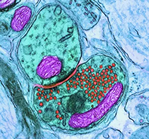
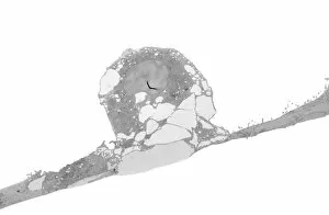
Background imageTransmission Electron Collection: Synapse nerve junction, TEM

Synapse nerve junction, TEM
Synapse. Coloured transmission electron micrograph (TEM) of a synapse, a junction between two nerve cells, in the brain. At a synapse an electrical signal is transmitted from one cell to the next in

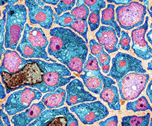
Background imageTransmission Electron Collection: Myelination of nerve fibres, TEM

Myelination of nerve fibres, TEM
Myelination of nerve fibres. Coloured transmission electron micrograph (TEM) of Schwann cells (blue, with brown nuclei) insulating nerve fibres (axons, pink) with a myelin sheath

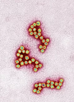
Background imageTransmission Electron Collection: Norovirus particles, TEM

Norovirus particles, TEM
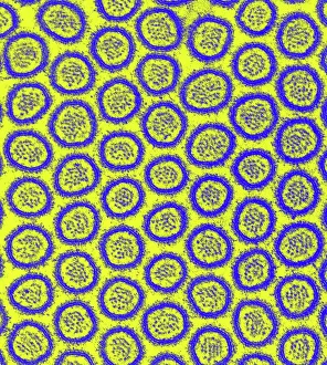
Norovirus particles. Coloured transmission electron micrograph (TEM) of norovirus particles. Norovirus is a genus of RNA (ribonucleic acid)

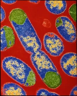
Background imageTransmission Electron Collection: E. coli bacteria

E. coli bacteria
False-colour transmission electron micrograph of the bacterium Escherichia coli, a normal inhabitant of the human intestine

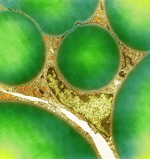
Background imageTransmission Electron Collection: Fat cells, TEM

Fat cells, TEM
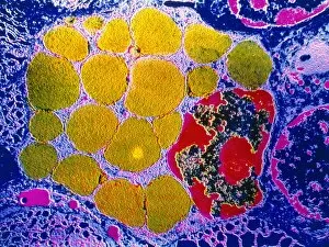
Fat cells. Coloured transmission electron micrograph (TEM) of a section through fat (adipose) tissue, consisting of fat-storing cells (adipocytes, green) supported by connective tissue

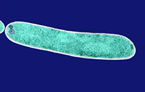
Background imageTransmission Electron Collection: E. coli bacterium, TEM

E. coli bacterium, TEM
E. coli bacterium, coloured transmission electron micrograph (TEM). E. coli are Gram-negative rod-shaped bacteria that are part of the normal flora of the human gut

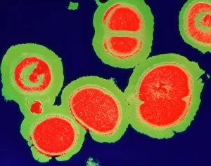
Background imageTransmission Electron Collection: MRSA resistant Staphylococcus bacteria

MRSA resistant Staphylococcus bacteria
MRSA: resistant Staphylococcus bacteria. Coloured transmission electron micrograph (TEM) of a deadly cluster of MRSA Staphylococcus aureus bacteria

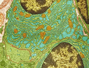
Background imageTransmission Electron Collection: Plasma cells, TEM

Plasma cells, TEM
Plasma cells. Coloured transmission electron micrograph (TEM) of plasma cells. Plasma cells are mature B lymphocytes (white blood cells) that produce and secrete antibodies during an immune response

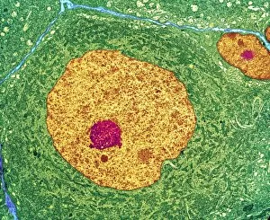
Background imageTransmission Electron Collection: Nerve cell, TEM

Nerve cell, TEM
Nerve cell. Coloured transmission electron micrograph (TEM) of a nerve cell body in cross- section. The cell has a large nucleus (yellow) and inner nucleolus (red)
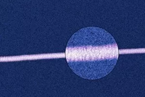
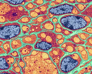
Background imageTransmission Electron Collection: Myelination of nerve fibres, TEM

Myelination of nerve fibres, TEM
Myelination of nerve fibres. Coloured transmission electron micrograph (TEM) of Schwann cells (red, with blue nuclei) insulating nerve fibres (axons, orange) with a myelin sheath

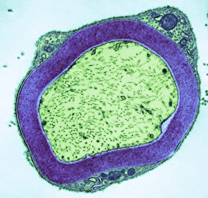
Background imageTransmission Electron Collection: Myelinated nerve, TEM

Myelinated nerve, TEM
Myelinated nerve. Coloured transmission electron micrograph (TEM) of myelinated nerve fibres and Schwann cells. Myelin (purple) is an insulating fatty layer that surrounds nerve fibres (axons)

Background imageTransmission Electron Collection: Mitochondrion, TEM

Mitochondrion, TEM
Mitochondrion. Coloured transmission electron micrograph (TEM) of mitochondria (green/blue) in heart muscle. Mitochondria are organelles found in the cytoplasm of eukaryotic cells

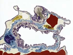
Background imageTransmission Electron Collection: Lung tissue, TEM

Lung tissue, TEM
Lung tissue, coloured transmission electron micrograph (TEM). A single alveolus is at center. At top right is a pulmonary capillary containing a red blood cell (red) and a platelet (yellow)

Background imageTransmission Electron Collection: Influenza virus particles, TEM

Influenza virus particles, TEM
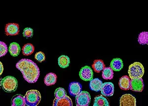
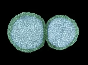
Influenza virus particles, coloured transmission electron micrograph (TEM). This virus is the cause of the respiratory disease influenza (flu) in humans and other animals

Background imageTransmission Electron Collection: Hepatitis C viruses, TEM

Hepatitis C viruses, TEM
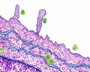
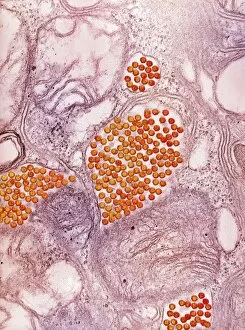
Hepatitis C viruses. Coloured transmission electron micrograph (TEM) of hepatitis C virus particles (green) infecting cultured liver cells (purple)

Background imageTransmission Electron Collection: Collagen fibres

Collagen fibres. Coloured transmission electron micrograph (TEM) of a section through healthy collagen fibres from human skin

Background imageTransmission Electron Collection: Intestinal microvilli, TEM

Intestinal microvilli, TEM
Intestinal microvilli, coloured transmission electron micrograph (TEM). The microvilli are finger-like projections (seen here in circular cross-section)

Background imageTransmission Electron Collection: E. coli bacterium, TEM

E. coli bacterium, TEM
E. coli bacterium. Coloured transmission electron micrograph (TEM) of an Escherichia coli bacterium in the early stages of binary fission, the process by which the bacterium divides

Background imageTransmission Electron Collection: HIV infected macrophage, SEM C018 / 8598

HIV infected macrophage, SEM C018 / 8598
HIV infected macrophage. Coloured ion-abrasion scanning electron micrograph (IA-SEM) of a macrophage white blood cell infected with human immunodeficiency virus (HIV, red)

Background imageTransmission Electron Collection: Macrophage and lymphocytes, TEM

Macrophage and lymphocytes, TEM
Macrophage and lymphocytes, transmission electron micrograph (TEM). Macrophage surrounded by lymphocytes in a lymph node. This view includes many intercellular contacts

Background imageTransmission Electron Collection: Influenza virus particles, TEM

Influenza virus particles, TEM
Influenza virus particles, coloured transmission electron micrograph (TEM). The virus consists of ribonucleic acid (RNA), surrounded by a nucleocapsid (blue) and a lipid envelope (green)

Background imageTransmission Electron Collection: Golgi apparatus, TEM C017 / 8234

Golgi apparatus, TEM C017 / 8234
Golgi apparatus. Transmission electron micrograph (TEM) of Golgi apparatus from a Chlamydomonas sp. alga. Golgi are membrane-bound organelles that modify and package proteins

Background imageTransmission Electron Collection: Skeletal muscle, TEM C016 / 5369

Skeletal muscle, TEM C016 / 5369
Skeletal muscle. Transmission electron micrograph (TEM) of a cross section through human skeletal (striated) muscle. Blocks of muscle (lighter grey) are surrounded by connective tissue (black)

Background imageTransmission Electron Collection: Cardiac muscle, TEM

Cardiac muscle, TEM
Cardiac muscle. Coloured transmission electron micrograph (TEM) of cardiac muscle fibrils (orange)from a healthy heart. Mitochondria (pink) supply the muscle cells with energy

Background imageTransmission Electron Collection: Cardiac muscle and capillary, TEM

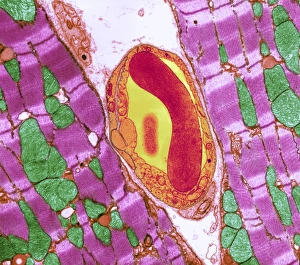
Cardiac muscle and capillary, TEM
Cardiac muscle. Coloured transmission electron micrograph (TEM) of cardiac muscle fibrils (purple)from a healthy heart. Mitochondria (green) supply the muscle cells with energy

Background imageTransmission Electron Collection: Mitochondria, TEM

Mitochondria, TEM
Mitochondria. Coloured transmission electron micrograph (TEM) of mitochondria (red) in heart muscle. Mitochondria are organelles found in the cytoplasm of eukaryotic cells

Background imageTransmission Electron Collection: Smallpox virus particle, TEM

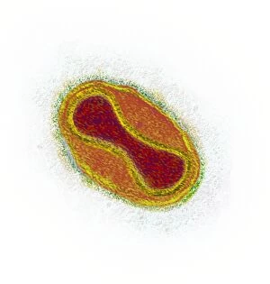
Smallpox virus particle, TEM
Smallpox virus particle. Coloured transmission electron micrograph (TEM) of a Variola major virus particle. Each particle consists of a DNA (deoxyribonucleic acid)

Background imageTransmission Electron Collection: Infectious bronchitis virus (IBV), TEM

Infectious bronchitis virus (IBV), TEM
Infectious bronchitis virus (IBV) particles, coloured transmission electron micrograph (TEM). This virus is a member of the coronavirus family

Background imageTransmission Electron Collection: Eastern equine encephalitis virus, TEM

Eastern equine encephalitis virus, TEM
Eastern equine encephalitis virus. Coloured transmission electron micrograph (TEM) of eastern equine encephalitis (EEE) virus particles (Alphavirus sp. orange) in the salivary gland of a mosquito

Background imageTransmission Electron Collection: HIV particles, TEM

HIV particles, TEM
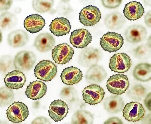
HIV (human immunodeficiency virus) particles, coloured transmission electron micrograph (TEM). Each particle consists of an RNA (ribonucleic acid, purple/brown) genome

Background imageTransmission Electron Collection: Yeast cells, TEM

Yeast cells, TEM
Yeast cells, coloured transmission electron micrograph (TEM). The cell wall is shown in blue, cytoplasm in red, and vacuole in green. Yeast is a single-celled fungus that reproduces asexually

Background imageTransmission Electron Collection: H1N1 swine flu virus, TEM C018 / 8602

H1N1 swine flu virus, TEM C018 / 8602
H1N1 swine flu virus. Coloured transmission electron micrograph (TEM) of swine influenza (flu) virus particles (virions, yellow)

Background imageTransmission Electron Collection: Brain nerve cells, TEM C014 / 0357

Brain nerve cells, TEM C014 / 0357
Brain nerve cells. Transmission electron micrograph (TEM) of a section through brain tissue from the cerebral cortex, showing numerous neurons (nerve cells) surrounded by axons and dendrites

Background imageTransmission Electron Collection: High-contrast direct DNA image, TEM

High-contrast direct DNA image, TEM
High-contrast direct DNA image. Coloured transmission electron micrograph (TEM) of the first high-contrast direct image of a bundle (fibre) of strands of DNA (deoxyribonucleic acid)

Background imageTransmission Electron Collection: Schmallenberg virus particles, TEM

Schmallenberg virus particles, TEM
Schmallenberg virus. Transmission electron micrograph (TEM) of virus particles (virions, yellow) of the Schmallenberg virus

Background imageTransmission Electron Collection: Activated plasma cell, TEM

Activated plasma cell, TEM
Activated plasma cell, coloured transmission electron micrograph (TEM). Plasma cells are derived from stimulated B lymphocytes (white blood cells)

Background imageTransmission Electron Collection: MERS coronavirus, TEM C015 / 1772

MERS coronavirus, TEM C015 / 1772
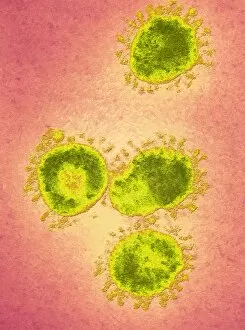
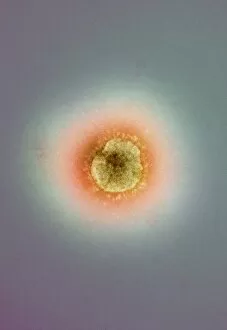
MERS coronavirus, coloured transmission electron micrograph (TEM). This virus (originally novel coronavirus 2012) has been named Middle East respiratory syndrome (MERS) coronavirus

Background imageTransmission Electron Collection: MERS coronavirus, TEM C015 / 1774

MERS coronavirus, TEM C015 / 1774
MERS coronavirus, coloured transmission electron micrograph (TEM). This virus (originally novel coronavirus 2012) has been named Middle East respiratory syndrome (MERS) coronavirus

Background imageTransmission Electron Collection: Granulocyte white blood cell, TEM

Granulocyte white blood cell, TEM
Granulocyte white blood cell. Coloured transmission electron micrograph (TEM) of a neutrophil granulocyte, a type of white blood cell

Background imageTransmission Electron Collection: MERS coronavirus, TEM C015 / 1776

MERS coronavirus, TEM C015 / 1776
MERS coronavirus, coloured transmission electron micrograph (TEM). This virus (originally novel coronavirus 2012) has been named Middle East respiratory syndrome (MERS) coronavirus

Background imageTransmission Electron Collection: MERS coronavirus, TEM C015 / 1773

MERS coronavirus, TEM C015 / 1773
MERS coronavirus, coloured transmission electron micrograph (TEM). This virus (originally novel coronavirus 2012) has been named Middle East respiratory syndrome (MERS) coronavirus

Background imageTransmission Electron Collection: Chloroplast, TEM C016 / 6297

Chloroplast, TEM C016 / 6297
Chloroplast. Coloured transmission electron micrograph (TEM) of chloroplast from the moss Physcomitrella patens. Chloroplasts are the sites of photosynthesis

Background imageTransmission Electron Collection: Chloroplast, TEM C016 / 6298

Chloroplast, TEM C016 / 6298
Chloroplast. Coloured transmission electron micrograph (TEM) of chloroplast from the moss Physcomitrella patens. Chloroplasts are the sites of photosynthesis

Background imageTransmission Electron Collection: Measles virus particle, TEM C015 / 7160

Measles virus particle, TEM C015 / 7160
Measles virus particle, coloured transmission electron micrograph (TEM). This virus, from the Morbillivirus group of viruses, consists of an RNA (ribonucleic acid)

Background imageTransmission Electron Collection: Measles virus particle, TEM C015 / 7161

Measles virus particle, TEM C015 / 7161
Measles virus particle, coloured transmission electron micrograph (TEM). This virus, from the Morbillivirus group of viruses, consists of an RNA (ribonucleic acid)

Background imageTransmission Electron Collection: MERS coronavirus, TEM C015 / 7155

MERS coronavirus, TEM C015 / 7155
MERS coronavirus. Coloured transmission electron micrograph (TEM) of MERS coronavirus particles (orange) budding from a host cell (brown)

Background imageTransmission Electron Collection: MERS coronavirus, TEM C015 / 7157

MERS coronavirus, TEM C015 / 7157
MERS coronavirus. Coloured transmission electron micrograph (TEM) of MERS coronavirus particles (small green dots) budding from a host cell (brown)

Background imageTransmission Electron Collection: MERS coronavirus, TEM C015 / 7158

MERS coronavirus, TEM C015 / 7158
MERS coronavirus. Transmission electron micrograph (TEM) of MERS coronavirus particles (small dots) budding from a host cell (centre)


All Professionally Made to Order for Quick Shipping

"Unlocking the Intricacies of Life: Exploring the Microscopic World through Transmission Electron Microscopy (TEM)" Delving into the depths of cellular structures and biological processes, transmission electron microscopy (TEM) has revolutionized our understanding of various aspects of life. With its high-resolution imaging capabilities, TEM allows us to witness intricate details that were once hidden from our sight. At synapse nerve junctions, TEM reveals a mesmerizing dance between neurotransmitters, enabling communication between neurons with remarkable precision. Norovirus particles come alive under TEM's lens, showcasing their unique shape and arrangement – a crucial insight in combating these notorious pathogens. E. Coli bacteria appear as tiny rods when observed through TEM, reminding us of their omnipresence in nature and sometimes unfortunate encounters in human health. Fat cells take on an unexpected beauty when magnified by TEM; their delicate structure resembling a web-like network that stores energy for our bodies. The myelination process is brought to life through TEM images capturing nerve fibers coated with protective sheaths. These stunning visuals help unravel the mysteries behind efficient neural signaling and hold promise for treating demyelinating diseases such as multiple sclerosis. TEM exposes the menacing presence of MRSA-resistant Staphylococcus bacteria – formidable adversaries in healthcare settings worldwide. Another glimpse at E. coli bacterium showcases its intricate internal machinery responsible for vital functions within this single-celled organism. Nerve cells reveal their complexity under TEM's scrutiny – branching dendrites reaching out like tree branches while axons transmit electrical signals across vast distances. Mitochondria steal the spotlight as they power these nerve cells' activities, appearing as dynamic organelles teeming with energy-producing potential. Plasma cells burst forth with vibrant colors when examined using TEM; their role in producing antibodies becomes even more awe-inspiring upon closer inspection. Once again, myelinated nerve fibers captivate us with their elegant architecture – a testament to nature's ingenuity in optimizing neural communication.