
Thoracic Vertebrae Collection
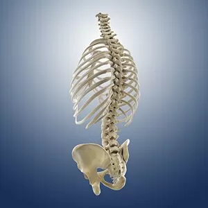
The thoracic vertebrae, also known as the upper back or middle back, play a crucial role in supporting and protecting our vital organs


All Professionally Made to Order for Quick Shipping
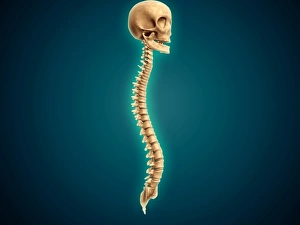
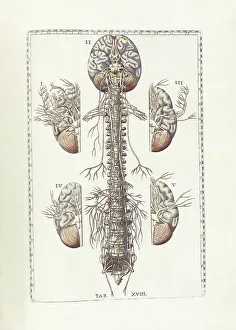
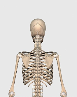
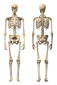
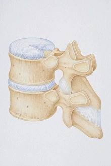
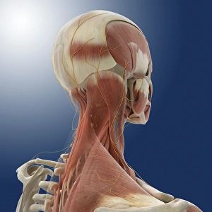
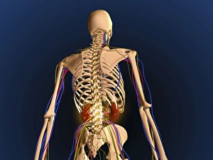
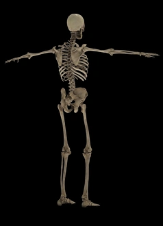
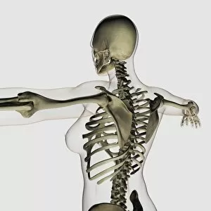
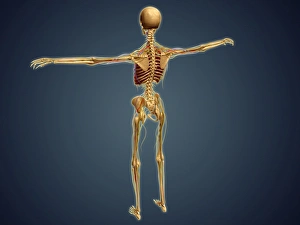
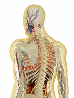
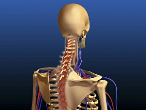
The thoracic vertebrae, also known as the upper back or middle back, play a crucial role in supporting and protecting our vital organs. As seen in the conceptual image of a human skull and spinal cord, these vertebrae form part of the intricate structure that connects our brain to the rest of our body. In Bartholomeo Eustachi's renowned work "The Science of Human Anatomy, " we can explore detailed illustrations showcasing the complexity and beauty of this region. From a rear view perspective, we witness the strength and elegance of the human skeletal system, with each thoracic vertebra contributing to its overall stability. Examining both front and back views of male human skeletons further emphasizes how these vertebrae fit into the larger framework. A diagram highlights essential components such as intervertebral disks, vertebral processes, and ligaments that ensure proper movement while maintaining structural integrity. Through a mesmerizing 3D rendering of the entire vertebral column accompanied by a skull, we gain an appreciation for how seamlessly these elements align to create harmony within our bodies. The side view showcases not only the spine but also provides insight into neck muscles and nerves' intricate network. As we zoom out from isolated images towards comprehensive depictions like those showing arteries, veins, nervous systems alongside skeletal structures—such as in artwork displaying a complete human body—we realize how interconnected everything truly is. This holistic approach allows us to understand why issues like inflammation in specific areas can impact multiple systems simultaneously. Ultimately captured through captivating conceptual imagery depicting just a backbone or medical illustrations highlighting inflammation concerns specifically within this region—the importance becomes evident. These remarkable bones serve as pillars upon which countless bodily functions rely—a testament to their significance within our complex anatomy.