Fraud Blocker Skip to main content

Thigh Bone Collection (page 4)

"The Thigh Bone: A Journey of Art, Replacement, and Resilience" Hip replacement meets artistry - a fusion of medical innovation and creative expression

Background imageThigh Bone Collection: Healthy pelvis, 3D CT scan

Healthy pelvis, 3D CT scan
Coloured three-dimensional (3D) computed tomography (CT) scan in front view of the healthy pelvis in a 52 year old patient showing the sacrum of the spine, pelvic bones

Background imageThigh Bone Collection: Growth disorder of thigh bone, X-ray

Growth disorder of thigh bone, X-ray
X-ray (front view) of the pelvis in an 8 year old patient showing ossification nuclei in the head of the thigh bone (femur, at left) due to the bone growth disorder chondrodysplasia punctata

Background imageThigh Bone Collection: Knee in anterior view, artwork

Knee in anterior view, artwork
Artwork of the anatomy of the knee joint (anterior view) showing the femur (thigh bone, at top) articulating with the tibia (shin bone, at bottom), cartilage (light blue)

Background imageThigh Bone Collection: Knee joint anatomy, artwork

Knee joint anatomy, artwork
Artwork of the anatomy of the knee joint (anterior view) showing the femur (thigh bone, at top) articulating with the tibia (shin bone, at bottom), articular cartilage

Background imageThigh Bone Collection: Slipped end of thigh bone, X-ray

Slipped end of thigh bone, X-ray
Coloured X-rays of the right hip of a young patient showing the slipped end (epiphysis) of the thigh bone (femur). During childhood the ends of the long bones are busy growing

Background imageThigh Bone Collection: Atherosclerosis in femoral arteries X-ray

Atherosclerosis in femoral arteries X-ray
Coloured X-ray (front view) of the pelvic region in a 72 year old patient showing atherosclerosis (mottled appearance) in the femoral arteries running on the inside of the femur (thigh) bones

Background imageThigh Bone Collection: Rheumatoid arthritis of the hip, X-ray

Rheumatoid arthritis of the hip, X-ray
Coloured X-ray (front view) of the pelvis in a 58 year old patient showing rheumatoid arthritis of the hip joint (at right)

Background imageThigh Bone Collection: Broken hip, X-ray

Broken hip, X-ray

Background imageThigh Bone Collection: Fractured pelvis, X-ray

Fractured pelvis, X-ray
Fractured pelvis. Coloured X-ray of fractures (red) in the pelvis. The head of the femur (thigh bone) articulates with the acetabulum to form the hip joint

Background imageThigh Bone Collection: Normal knee, X-ray

Normal knee, X-ray
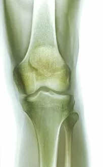
Normal knee, coloured frontal X-ray. The tibia (shin bone, bottom centre) and the smaller fibula (bottom right) articulate with the femur (thigh bone, top) to form the knee joint

Background imageThigh Bone Collection: Fractured femur, X-ray

Fractured femur, X-ray
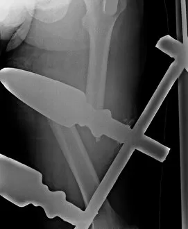
Fractured femur. X-ray of a patient with a total hip replacement who has fractured their femur (thigh bone). The fracture (centre right) has occurred towards the end of the implant (solid white)

Background imageThigh Bone Collection: Complete thigh-bone fracture, X-ray

Complete thigh-bone fracture, X-ray
X-ray (back view) of the leg of an adult patient, showing a complete fracture of the thigh bone (femur, running top centre to bottom)

Background imageThigh Bone Collection: Neck of femur fracture, X-ray

Neck of femur fracture, X-ray
Coloured X-ray (front view) of the pelvis of a patient, showing a hip fracture (at left) to the neck of the thigh bone (femur)


All Professionally Made to Order for Quick Shipping

"The Thigh Bone: A Journey of Art, Replacement, and Resilience" Hip replacement meets artistry - a fusion of medical innovation and creative expression. Peering into the depths of healing - an X-ray reveals the success of total hip replacement surgery. From injury to inspiration - damaged knee ligament transformed into captivating artwork. Unveiling strength within - a healthy knee captured in its intricate beauty through an X-ray lens. Exploring the human frame - an enchanting engraving depicting the timeless elegance of our skeleton. Beneath the surface lies perfection - normal legs revealed through mesmerizing X-rays. Literary musings on anatomy's marvels - Henry Fielding's illustration brings Tom Jones to life alongside a femur bone engraving. Delving deeper for answers – CT scan captures the essence of a healthy knee in stunning detail (CT scan C018 / 0413). Celebrating power from behind – artistic portrayal showcases buttock muscles' strength and grace (artwork C013 / 4414). The foundation of our being unveiled – an exquisite illustration showcasing the majesty of human bones, including the mighty femur. Culinary exploration meets anatomy – using a kitchen knife to remove bone from chicken thigh highlights similarities between nature and ourselves. Osteoarthritis unraveled–an X-ray revealing F006/3744 showcases how it affects one's hip joint.