
Temporal Lobe Collection
The temporal lobe, a fascinating region of the human brain, is often overlooked but plays a crucial role in our daily lives


All Professionally Made to Order for Quick Shipping
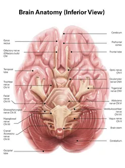
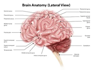
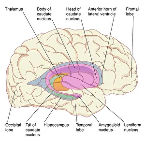
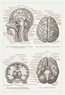
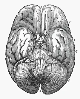
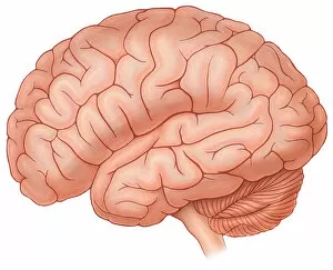
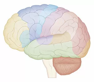
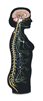
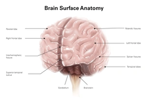
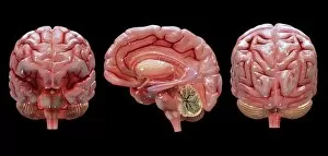
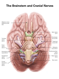
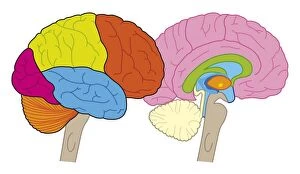
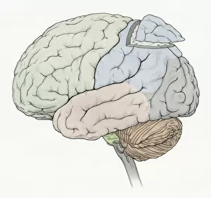
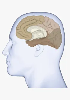
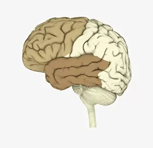
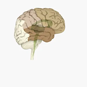
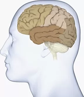
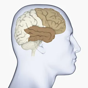
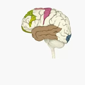
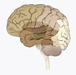
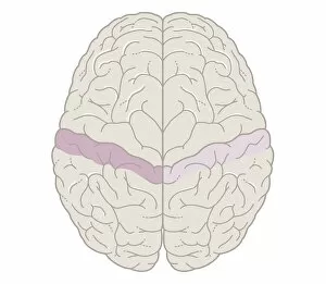
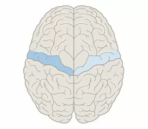
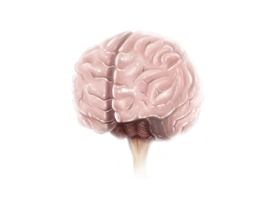
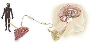
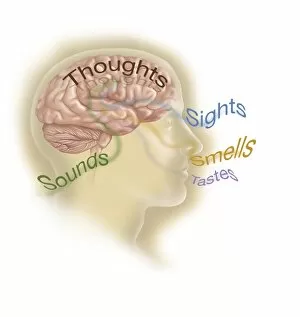
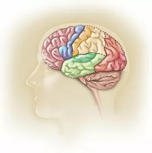
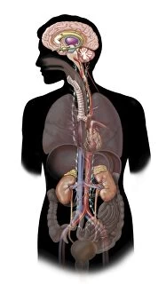
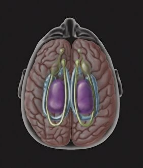
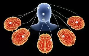
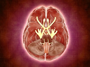
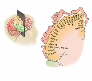
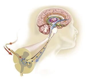
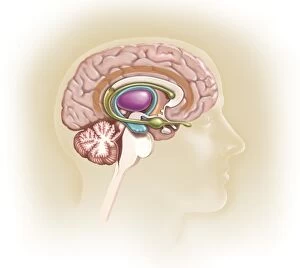
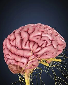
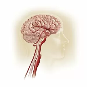
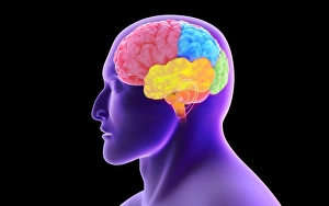
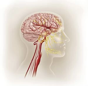
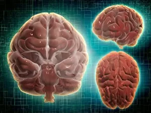
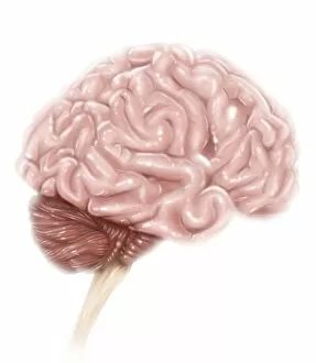
The temporal lobe, a fascinating region of the human brain, is often overlooked but plays a crucial role in our daily lives. When we examine its anatomy from an inferior view, we can appreciate its intricate structure and connections within the vast network of our minds. In lateral view, this remarkable part of the brain reveals itself alongside other lobes, showcasing its unique features. Artwork depicting the basal ganglia further emphasizes the significance of the temporal lobe's functions. Dating back to 1876, a lithograph beautifully captures its position and importance within the overall architecture of our brains. Similarly, an engraved illustration from circa 1880 showcases the under surface of this extraordinary organ. A cross-sectional biomedical illustration provides us with a detailed map that highlights how different areas within this lobe contribute to various cognitive processes. As we explore deeper into brain anatomy, we discover how it interacts with both autonomic nervous system and limbic system in regulating emotions and bodily functions. Brain surface anatomy illustrations with labels enable us to identify specific structures associated with the temporal lobe accurately. This side view also allows us to understand how it fits harmoniously among other functional lobes responsible for distinct aspects of cognition. In modern times, digital illustrations provide even more clarity as they showcase not only individual lobes but also cross-sections that reveal their inner workings. These visuals help us comprehend how frontal, occipital lobes coexist alongside sections removed from parietal lobe while highlighting vital components like cerebellum and medulla oblongata. Overall, exploring these captivating depictions enables us to grasp just how essential the temporal lobe is in shaping who we are as individuals. Its involvement in memory formation, language processing, auditory perception makes it truly remarkable - reminding us once again why understanding our own brains remains an endless source of fascination for humanity throughout history.