Fraud Blocker Skip to main content

Tb Collection (page 5)

"Unveiling the Historical Battle Against TB

Background imageTb Collection: Macrophage engulfing TB bacteria, SEM C017 / 8309

Macrophage engulfing TB bacteria, SEM C017 / 8309
Macrophage engulfing TB bacteria. Coloured scanning electron micrograph (SEM) of a macrophage white blood cell engulfing tuberculosis (Mycobacterium tuberculosis) bacteria (pink)

Background imageTb Collection: Tuberculosis campaign, early 20th century C017 / 7885

Tuberculosis campaign, early 20th century C017 / 7885
Tuberculosis campaign, early 20th century. Doctor using a stethoscope to check a boys lungs for signs of the respiratory bacterial disease tuberculosis (TB)

Background imageTb Collection: Tuberculosis, light micrograph C015 / 6400

Tuberculosis, light micrograph C015 / 6400
Tuberculosis. Light micrograph of a section through a lymph node showing a Langhans cell (centre) caused by tuberculosis (TB)

Background imageTb Collection: Spleen tuberculosis C015 / 6399

Spleen tuberculosis C015 / 6399
Spleen tuberculosis. Gross specimen of a sectioned spleen from a patient with miliary tuberculosis (TB). Tubercules, nodular lesions of infected dead tissue, are white

Background imageTb Collection: Wife of unemployed coal miner, suffering from TB, living in

Wife of unemployed coal miner, suffering from TB, living in old company store. Abandoned mining town of Marine, West Virginia. (See 50110-D)J. Date 1938 Sept

Background imageTb Collection: Dagshai Parade Ground

Dagshai Parade Ground - Built by the British as a sanatorium for TB patients in Himachal Pradesh State, Solan District. In 1920 there was a large-scale mutiny by Irish soldiers stationed at Dagsai

Background imageTb Collection: Edward VII Memorial Hospital, Machynlleth

Edward VII Memorial Hospital, Machynlleth
The Edward VII Memorial Hospital at Machynlleth, Montgomeryshire. The hospital, located in the former Machynlleth Union workhouse, opened in 1920 for the treatment of tuberculosis (TB) patients

Background imageTb Collection: Central London Sick Asylum, Hendon

Central London Sick Asylum, Hendon
Interior of the Central London Sick Asylum opened in 1898 at Hendon, Middlesex. Later known as Hendon Infirmary and Colindale Hospital, it was a treatment centre for tuberculosis (TB) patients

Background imageTb Collection: Testing sputum for lung diseases

Testing sputum for lung diseases. Image 3 of 3. Researcher applying sputum (mucus coughed up from the lungs) from a patient to a glass slide

Background imageTb Collection: Lung scarring from tuberculosis, X-ray

Lung scarring from tuberculosis, X-ray
Tuberculosis lung scarring. Coloured chest X-ray showing scarring (green) of the lungs after a case of chronic tuberculosis (TB). The heart (red) is also seen

Background imageTb Collection: Potts disease

Potts disease. Coloured X-ray of the spine of a patient with Potts disease, or tuberculosis (TB) of the spine. The vertebrae (brown blocks) have become compacted and inflamed (red, yellow)

Background imageTb Collection: Macrophages and tuberculosis vaccine

Macrophages and tuberculosis vaccine. Fluorescence light micrograph of macrophage (red, outlined in black) white blood cells that have phagocytosed (engulfed) Mycobacterium bovis bacteria (pink)

Background imageTb Collection: Macrophage and tuberculosis vaccine, TEM

Macrophage and tuberculosis vaccine, TEM
Macrophage and tuberculosis vaccine. Coloured transmission electron micrograph (TEM) of phagocytosed (engulfed) Mycobacterium bovis bacteria (purple) in a macrophage (green) white blood cell

Background imageTb Collection: Macrophage engulfing bacteria, SEM

Macrophage engulfing bacteria, SEM
Macrophage engulfing bacteria. Coloured scanning electron micrograph (SEM) of a macrophage white blood cell (brown) engulfing Mycobacterium tuberculosis bacteria (yellow)

Background imageTb Collection: Macrophage engulfing tuberculosis vaccine

Macrophage engulfing tuberculosis vaccine. Coloured scanning electron micrograph (SEM) of a macrophage white blood cell engulfing Mycobacterium bovis bacteria (yellow)

Background imageTb Collection: Liver tuberculosis, light micrograph

Liver tuberculosis, light micrograph
Liver tuberculosis. Coloured light micrograph of a section through the liver of a patient with miliary tuberculosis (TB). A tubercle, a nodular lesion of infected dead tissue, is seen at left

Background imageTb Collection: Miliary tuberculosis, light micrograph

Miliary tuberculosis, light micrograph
Miliary tuberculosis. Light micrograph of a section through a single miliary tubercle (left) in a patients lung. Tubercles are nodular lesions of infected dead tissue that arise from tuberculosis (TB)

Background imageTb Collection: Tuberculosis resistance to some drugs

Tuberculosis resistance to some drugs
Conceptual computer illustration showing the virtual brick wall resistance of Tuberculosis (TB) to drugs. Drug resistance is the reduction in effectiveness of a drug such as an antimicrobial or an

Background imageTb Collection: Illustration of E. coli in a Petri dish

Illustration of E. coli in a Petri dish
Conceptual computer illustration of E.coli bacteria being cultured in a Petri dish. Escherichia coli has the ability to ferment carbohydrates

Background imageTb Collection: Tuberculosis bacteria, artwork

Tuberculosis bacteria, artwork

Background imageTb Collection: Koch and tuberculosis, 19th century

Koch and tuberculosis, 19th century
Koch and tuberculosis research. The German bacteriologist Robert Koch (1843-1910) is considered one of the founders of modern medical bacteriology

Background imageTb Collection: TB bacteria infecting macrophages, SEM

TB bacteria infecting macrophages, SEM
TB bacteria infecting macrophages. Coloured scanning electron micrograph (SEM) of Mycobacterium tuberculosis bacteria (orange) invading macrophage white blood cells (pink)

Background imageTb Collection: Terbium

Terbium. Sample of the rare earth metal Terbium (Tb). The rare earth metals, or rare earth elements, are a set of seventeen chemical elements in the periodic table

Background imageTb Collection: Tuberculosis diagnosis, 19th century

Tuberculosis diagnosis, 19th century. Artwork showing the French physician Rene Laennec (1781-1826), at the Necker Hospital, Paris, France, examining a patient (left)

Background imageTb Collection: Neutrophil engulfing fungus, SEM

Neutrophil engulfing fungus, SEM
Neutrophil engulfing fungus. Coloured scanning electron micrograph (SEM) of a neutrophil white blood cell (orange) engulfing and destroying a Candida albicans (thrush) fungal hypha (green)

Background imageTb Collection: Biochip for detection of tuberculosis

Biochip for detection of tuberculosis
Biochip production. Close-up view of some TB-Biochips. The chips being made here are used to detect tuberculosis, particularly drug-resistant strains

Background imageTb Collection: Tuberculosis lymphadenopathy, X-ray

Tuberculosis lymphadenopathy, X-ray
Coloured X-ray (front view) of the chest in a 14 year old male patient, showing inflamed lymph nodes (lymphadenopathy, yellow, at centre right and left) caused by tuberculosis (TB)

Background imageTb Collection: Old and new tuberculosis, X-ray

Old and new tuberculosis, X-ray
Coloured X-ray (front view) of the chest in a 50 year old male patient, showing fibrosis in the lungs from old tuberculosis (more fibrous scarring, orange)

Background imageTb Collection: Neutrophil engulfing TB bacteria, SEM

Neutrophil engulfing TB bacteria, SEM
Neutrophil engulfing TB bacteria. Coloured scanning electron micrograph (SEM) of a neutrophil white blood cell (large, centre) engulfing and destroying a tuberculosis (Mycobacterium tuberculosis)

Background imageTb Collection: Neutrophil engulfing thrush fungus, SEM

Neutrophil engulfing thrush fungus, SEM
Neutrophil engulfing thrush fungus. Coloured scanning electron micrograph (SEM) of a neutrophil white blood cell engulfing and destroying a hypha (round)

Background imageTb Collection: Mycobacterium bovis, TEM

Mycobacterium bovis, TEM
Mycobacterium bovis. Coloured transmission electron micrograph (TEM) of a group of M. bovis bacteria. It is a Gram-positive, aerobic, non- motile bacterium. M. bovis is a variant of M

Background imageTb Collection: Tuberculosis vaccine bacteria, SEM

Tuberculosis vaccine bacteria, SEM
Tuberculosis vaccine bacteria. Coloured scanning electron micrograph (SEM) of the Gram-positive rod-shaped (bacillus) Mycobacterium bovis bacteria

Background imageTb Collection: Tuberculosis bacteria, TEM

Tuberculosis bacteria, TEM
Tuberculosis bacteria. Coloured transmission electron micrograph (TEM) of a section through Mycobacterium tuberculosis bacteria

Background imageTb Collection: Eurasian Badger (Meles meles) adult, emerging from sett, in daylight, Oxfordshire, England

Eurasian Badger (Meles meles) adult, emerging from sett, in daylight, Oxfordshire, England

Background imageTb Collection: Eurasian Badger (Meles meles) cub, foraging on sett, in daylight, Oxfordshire, England

Eurasian Badger (Meles meles) cub, foraging on sett, in daylight, Oxfordshire, England

Background imageTb Collection: Eurasian Badger (Meles meles) cub, foraging amongst vegetation, in daylight, Oxfordshire, England

Eurasian Badger (Meles meles) cub, foraging amongst vegetation, in daylight, Oxfordshire, England

Background imageTb Collection: Eurasian Badger (Meles meles) cub, on chalk spoil heap, in daylight, Oxfordshire, England

Eurasian Badger (Meles meles) cub, on chalk spoil heap, in daylight, Oxfordshire, England

Background imageTb Collection: Main entrance, Pinewood Sanatorium, Wokingham

Main entrance, Pinewood Sanatorium, Wokingham
View of the main entrance of Pinewood Sanatorium at Bagshot Sands, near Wokingham, Berkshire, originally opened in 1898 as the London Open Air Sanatorium by the National Association for

Background imageTb Collection: Gymnasium, Princess Marys Hospital, Margate, Kent

Gymnasium, Princess Marys Hospital, Margate, Kent
The gymnasium and physiotherapy department at Princess Marys Hospital, Margate, Kent. A wide range of equipment includes an exercise bicycle, a ships wheel, and part of a bus

Background imageTb Collection: Consumption cases, NIPRCC East Harling, Norfolk

Consumption cases, NIPRCC East Harling, Norfolk
Consumption (tuberculosis) cases lying out on a balcony at the East Harling, Norfolk, branch of the National Institutions for Persons Requiring Care and Control


All Professionally Made to Order for Quick Shipping

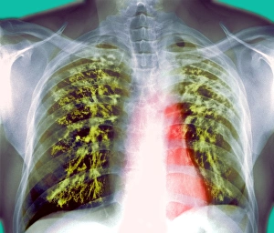
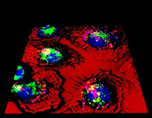
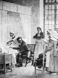
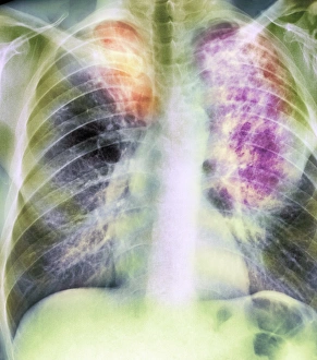
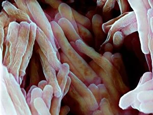
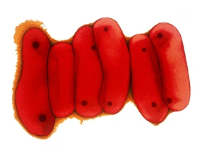
"Unveiling the Historical Battle Against TB: From Shotley Bridge General Hospital to Highwood Hospital" Step into the past as we journey through the fight against tuberculosis (TB), a disease that plagued communities for centuries. Our first stop takes us to Shotley Bridge General Hospital in County Durham, where medical professionals dedicated their efforts to treating TB patients. Moving on, we arrive at Highwood Hospital in Brentwood, Essex, witnessing groundbreaking advancements in Calots spinal surgery during the 19th century. This procedure aimed to alleviate complications caused by spinal tuberculosis and offered hope for those suffering from this debilitating condition. Fast forward to modern times, where X-ray technology revolutionized TB diagnosis. Witnessing an X-ray of Tuberculosis showcases how medical imaging became instrumental in identifying and monitoring this infectious disease. Shifting gears from medicine to sports, we find ourselves amidst The Eton vs. Harrow Cricket Match at Lords in 1910. Little did they know that even within these prestigious sporting events, individuals may have been silently battling with TB. Our journey continues as we explore Highwood School in Brentwood, Essex – a place where education thrived despite the presence of nearby Highwood Hospital. These institutions stood side by side as reminders of both resilience and vulnerability when it came to combating tuberculosis. Zooming into microscopic levels reveals bacteria infecting a macrophage under scanning electron microscopy (SEM). This captivating image highlights how TB infiltrates our immune system's defense mechanisms while providing valuable insights for researchers striving towards effective treatments. As our historical exploration nears its end, we visit Empire Hotel in Lowestoft, Suffolk – a coastal retreat often frequented by those seeking respite from urban environments affected by tuberculosis outbreaks. Here people could escape temporarily from the shadow cast by this relentless disease. Finally, another SEM image captures a macrophage engulfing TB bacteria—a visual representation of our immune system's ongoing battle against this persistent foe.