
Sweat Gland Collection
The sweat gland is a vital component of the human skin, playing a crucial role in maintaining our body's temperature and overall health


All Professionally Made to Order for Quick Shipping
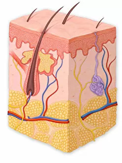
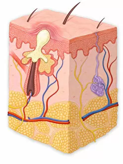
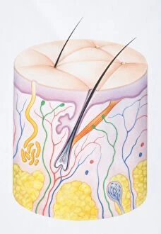
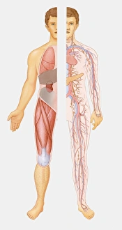
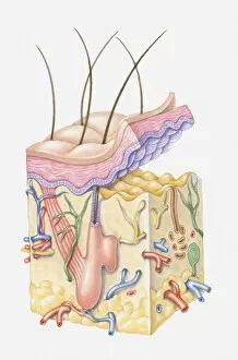
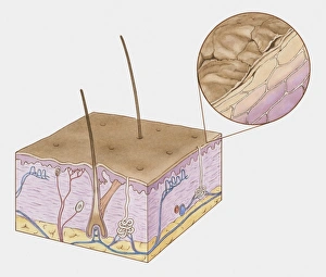
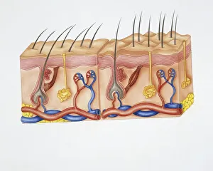
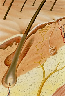
The sweat gland is a vital component of the human skin, playing a crucial role in maintaining our body's temperature and overall health. A normal cross section of the skin reveals its intricate layers, including the epidermis, dermis, and hair follicle. These layers work together to protect us from external threats while allowing essential functions to take place. In this detailed view of the skin cross section, we can observe various common dermatological issues. A blackhead appears as a clogged pore filled with excess oil and dead skin cells. It serves as a reminder to keep our pores clean through proper skincare routines. A papule is another type of blemish that arises when there is inflammation or infection within the skin. This small raised area may be red or pink in color and often requires targeted treatment for resolution. Pustules are similar to papules but contain pus due to an accumulation of white blood cells fighting off bacteria or other pathogens and can be seen as small bumps on the surface of the skin and should not be squeezed or popped without professional guidance. Whiteheads are yet another common occurrence in which blocked pores trap sebum beneath the surface of our skin. These closed comedones appear as tiny white bumps and necessitate gentle exfoliation techniques for their removal. An illustrative diagram showcases how these different components fit into our internal systems within the human body structure. Our intricate network consists not only of organs but also includes complex tissues like our remarkable integumentary system – comprising both hair and skin – which acts as an interface between us and our environment. This biomedical illustration provides a cross-sectional view highlighting each layer's unique characteristics within human skin structure: epidermis, dermis, hair follicles, sweat glands, nerve endings, sebaceous glands (responsible for producing sebum), and circulatory system vessels supplying nutrients throughout this dynamic organ. Underneath it all lies microscopic evidence revealing fascinating details about our skin's composition.