
Surgery Collection (page 2)
"Surgery: A Journey Through Time and Advancements in Medical Science" Step into the world of surgery


All Professionally Made to Order for Quick Shipping
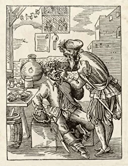
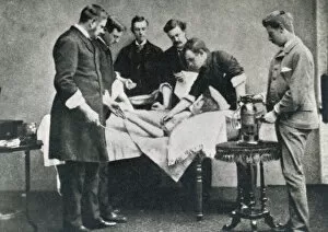
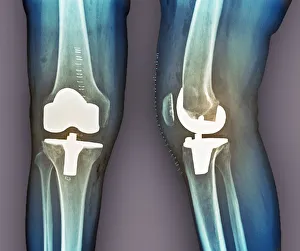
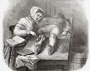
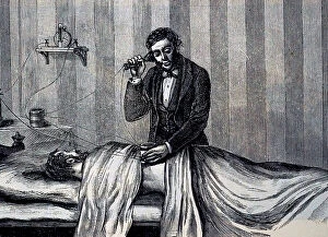
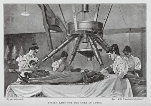
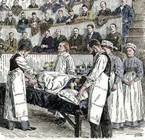
"Surgery: A Journey Through Time and Advancements in Medical Science" Step into the world of surgery, where history meets innovation and medical breakthroughs have transformed lives. In 1846, a monumental milestone was achieved with the first use of anesthesia in surgery, forever changing the way procedures were conducted. Gazing upon a Roman statue of Asclepius, the Greek god associated with healing and medicine, one can't help but marvel at how far we've come. From trepanation depicted in 14th-century artwork to evidence of trephination found in an Inca skull, ancient civilizations had their own methods for surgical interventions. The cause and effect relationship between dental health and overall well-being is humorously portrayed by H. M. Bateman's dentist cartoon. It reminds us that even seemingly simple procedures like tooth extraction required skilled professionals throughout history. Nurses diligently watch over surgeries as silent guardians of patient care. Their presence brings comfort amidst complex operations such as total hip replacements captured on X-rays or Calots spinal surgery from the 19th century. In 1947, the Medical Fund Society Dental Surgery provided much-needed oral healthcare to those who couldn't afford it. This initiative highlights society's commitment to ensuring access to essential medical services for all. Even armies recognized the importance of dental health with mobile dental surgeries like that belonging to the French army—an innovative approach bringing dentistry closer to soldiers during times of conflict. Amidst these advancements lies a reminder that surgery has not always been about progress alone; it also involved difficult decisions like amputations—whether it be an arm or leg—the ultimate sacrifice made for survival or quality of life. As we reflect on this captivating journey through time, let us appreciate how surgical techniques have evolved while honoring those who paved the way for modern medicine—a testament to human resilience and our unwavering pursuit of better healthcare outcomes.