mail_outline sales@mediastorehouse.com
Framed Print
Premium Framed Print
Canvas Print
Metal Print
Photographic Print
Poster Print
Fine Art Print
Jigsaw Puzzle
Photo Mug
Pillow
Mouse Mat
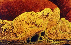
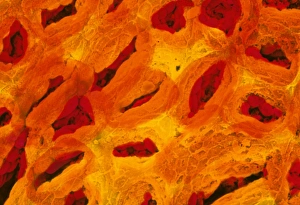
Colour SEM of cross-section through stomach wallStomach mucosa. Coloured Scanning Electron Micrograph (SEM) of a cross section through the mucous membrane (or mucosa) lining the inside of the human stomach. The mucosa is the thick yellow layer
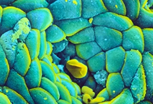
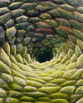
Coloured SEM of the stomach mucosa & gastric pitStomach wall. Coloured Scanning Electron Micrograph (SEM) of the surface of the fundus region of the human stomach. At centre is a dark opening known as a gastric pit
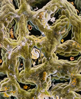
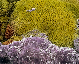
False-colour SEM of secreting gastric glandsFalse-colour scanning electron micrograph (SEM) of secreting gastric glands of the stomach. Gastric glands occur as downgrowths of the stomach epithe- lium, forming pits (seen here)
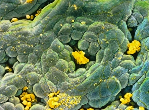
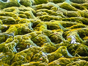
False-colour SEM of glandular stomach wallFalse-colour scanning electron micrograph (SEM) of the glandular lining of the stomach. The gastric mucosa is the site of production of hydrochloric acid, digestive enzymes, hormones and mucous
False-colour SEM of gastric gland in stomachGastric gland: false-colour scanning electron micrograph (SEM) of the mucous membrane lining the stomach, featuring a single, circular gastric gland
False colour SEM of entrance to gastric glandFalse-colour scanning electron micrograph (SEM) of the mucous membrane lining the stomach, showing the entrance to a gastric gland
F / colour SEM of oesophagus-stomach transition zoneF/colour SEM of oesophagus-stomach transition zone
False-colour SEM of gastric glands of stomachGastric gland: false-colour scanning electron micrograph (SEM) of the mucous membrane lining the stomach, showing the shadowy entrances to a number of gastric glands
Colour SEM of the stomach mucosa & gastric pits