
Staphylococcus Collection
Staphylococcus, a group of bacteria commonly found on the skin and mucous membranes of humans and animals, has both beneficial and harmful effects


All Professionally Made to Order for Quick Shipping
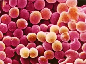
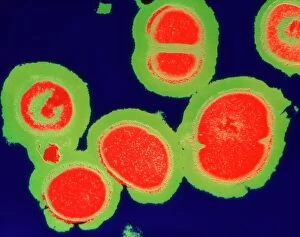
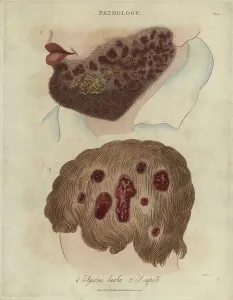
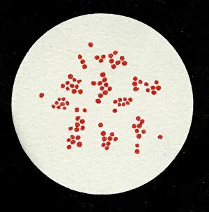
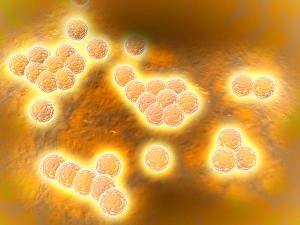
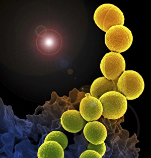
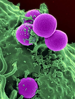
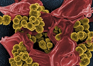
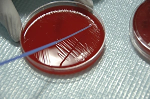
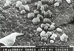
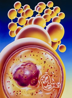
Staphylococcus, a group of bacteria commonly found on the skin and mucous membranes of humans and animals, has both beneficial and harmful effects. Staphylococcus aureus is one such bacterium that can cause various infections in humans. MRSA (methicillin-resistant Staphylococcus aureus) is a strain of this bacterium that has developed resistance to multiple antibiotics, making it difficult to treat. In 1906, a lithograph captured the moment when Staphylococcus pyogenes underwent cell division, showcasing its ability to multiply rapidly. Another lithograph from 1905 depicted a colony bacteria, highlighting their unique appearance under the microscope. Speaking of microscopic views, these images reveal the intricate structure bacteria as seen through powerful lenses. The vibrant colors bring attention to Methicillin-resistant Staphylococcus aureus (MRSA), emphasizing its significance in healthcare settings where it poses a serious threat. Interestingly, scanning electron micrographs showcase how our immune system fights against MRSA infection. A white blood cell can be observed devouring MRSA while human neutrophils are shown ingesting these resistant bacteria. Studying staphylococcus helps scientists understand its behavior and devise strategies for prevention and treatment. While some strains cause harm by triggering infections like pneumonia or skin abscesses, others play crucial roles in maintaining healthy skin flora.