
Sporangia Collection
Sporangia: Nature's Tiny Marvels In the enchanting woodlands of Buckinghamshire, England, a fascinating world unfolds


All Professionally Made to Order for Quick Shipping
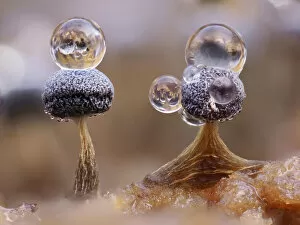
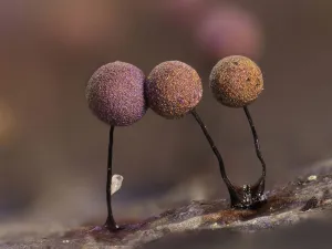
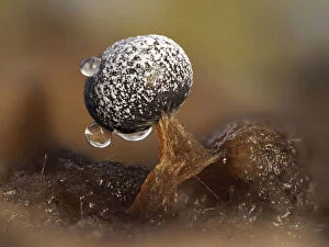
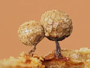
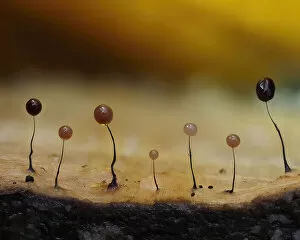
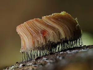
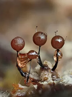
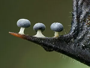
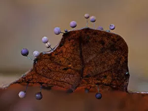
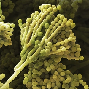
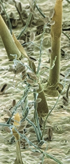
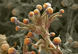
Sporangia: Nature's Tiny Marvels In the enchanting woodlands of Buckinghamshire, England, a fascinating world unfolds. Slime mould sporangia emerge from their hidden realms, captivating the eyes and minds of those fortunate enough to witness their beauty. Amongst the bark's edge, delicate slime mould (Stemonitopsis typhina) sporangia thrive in November. These tiny structures stand tall, focus stacked to reveal intricate details that would otherwise go unnoticed. A testament to nature's artistry. Metatrichia floriformis takes center stage in January as its line splits open with grace. Like a symphony reaching its crescendo, spores are released into the air, ready to embark on new journeys across Buckinghamshire's landscape. Lamproderma scintillans unveils its secrets through an astonishing close-up shot. Standing at a mere 1mm tall, these sporangia hold immense power within their minuscule frames. Their presence is ethereal yet undeniable. Physarum album showcases dew droplets delicately resting upon two sporangia in a mesmerizing close-up shot. The juxtaposition of water and life creates an image that evokes both tranquility and wonder. As September arrives in Hertfordshire, Stemonitis flavogenita begins its transformation from green to orange hues on an Oak log. These maturing sporangia symbolize growth and change amidst nature's cycle - a sight worth beholding. August brings Lamproderma arcyrionema into focus as three immature sporangia begin their journey towards maturity. Standing at just one millimeter tall, they represent resilience and determination against all odds. On decaying leaves lie two Lamproderma scintillans sporangia with peridium split open like doors inviting exploration into another realm entirely. February witnesses this magical scene unfold before our eyes, beckoning us to delve deeper into nature's mysteries.