Fraud Blocker Skip to main content

Sinuses Collection

"Exploring the Intricacies of Sinuses

Background imageSinuses Collection: Skull anatomy by Leonardo da Vinci

Skull anatomy by Leonardo da Vinci. Historical artwork and notes on the anatomy of the human skull and teeth, by the Italian artist and scientist Leonardo da Vinci (1452-1519)

Background imageSinuses Collection: Skull anatomy

Skull anatomy. Historical anatomical artwork of various views of the human skull. The frontal view (upper left) shows the teeth, nose and eye spaces

Background imageSinuses Collection: Nose and sinuses, CT scan

Nose and sinuses, CT scan
Nose anatomy and sinuses. Coloured computed tomography (CT) scan of a frontal view through the nose of a 31 year old woman

Background imageSinuses Collection: Sectional dissected head, 19th century C017 / 0705

Sectional dissected head, 19th century C017 / 0705
Dissected cross section of a human head and neck with the brain removed. Anatomy of the skull, spine, teeth and sinuses can be seen. This artefact dates from the nineteenth century

Background imageSinuses Collection: Brain sinuses, 1825 artwork

Brain sinuses, 1825 artwork
Brain sinuses. Axial section through the head at the level of the base of the skull (front of head at top), showing the sinuses (blue) of the brains dura mater

Background imageSinuses Collection: Lymph node, artwork C013 / 4632

Lymph node, artwork C013 / 4632
Lymph node. Computer artwork of a section through a human lymph node. Lymph nodes are kidney-shaped structures located at intervals along lymph vessels (tubes)

Background imageSinuses Collection: Lymph node, artwork C013 / 4631

Lymph node, artwork C013 / 4631
Lymph node. Computer artwork of a section through a human lymph node. Lymph nodes are kidney-shaped structures located at intervals along lymph vessels (tubes)

Background imageSinuses Collection: Nose, mouth and throat

Nose, mouth and throat. Cutaway artwork of the anatomy of the nose, mouth and throat. The large nasal cavity (upper centre) is lined with nerves (yellow) branching from the olfactory bulb (above)

Background imageSinuses Collection: Brain and arteries, coloured CT scan

Brain and arteries, coloured CT scan
Healthy brain and arteries. Coloured computed tomography (CT) scan showing healthy arteries (white) supplying a human brain (red) with oxygenated blood. The front of the head is at top

Background imageSinuses Collection: Healthy brain and frontal sinus, CT scan

Healthy brain and frontal sinus, CT scan
Healthy brain and frontal sinus. Coloured computed tomography (CT) scan of a human head. The axial (horizontal) scan has revealed the head and brain in cross section. The front of the head is at top

Background imageSinuses Collection: Healthy brain and eyes, CT scan

Healthy brain and eyes, CT scan
Healthy brain and eyes. Coloured computed tomography (CT) scan of a human head. The axial (horizontal) scan has revealed the head and brain in cross section at eye-level

Background imageSinuses Collection: Healthy brain, MRI scan

Healthy brain, MRI scan
Healthy brain, coloured magnetic resonance imaging (MRI) scan. The brain is seen in profile. The nose is at far right, and behind it are the sinuses (dark), air spaces in the skull

Background imageSinuses Collection: Healthy brain, CT scan

Healthy brain, CT scan
Healthy brain, coloured computed tomography (CT) scan. The front of the brain is at top. The brown circles are the eyeballs

Background imageSinuses Collection: Healthy skull, coloured X-ray

Healthy skull, coloured X-ray
Healthy human skull. Side view X-ray showing the skull and the outline of the head (black). The bones of the cranium are fused together to form a complete structure that protects the brain

Background imageSinuses Collection: Human skull, computer artwork

Human skull, computer artwork
Skull, computer artwork. The bones of the cranium (top) are fused together to form a solid structure that encloses and protects the brain. At bottom are the teeth, set in the jaw

Background imageSinuses Collection: Skull, 3-D CT scan

Skull, 3-D CT scan
Human skull, 3-D computed tomography (CT) scan. The orbits of the eyes are either side of the nasal cavity. The front of the skull contains large air spaces called sinuses

Background imageSinuses Collection: Tired woman

Tired woman
MODEL RELEASED. Tired woman rubbing her eye

Background imageSinuses Collection: Normal head and brain, MRI scan

Normal head and brain, MRI scan
Normal head and brain. Coloured profile (sagittal) magnetic resonance imaging (MRI) scan through the centre of the head and brain

Background imageSinuses Collection: Normal brain, CT scan

Normal brain, CT scan
Normal brain, coloured axial (cross-sectional) computed tomography (CT) scan. The front of the head is at top. The brain is yellow (lower centre)

Background imageSinuses Collection: Respiratory virus infection treatment

Respiratory virus infection treatment. Computer artwork showing virus particles (spherical) inside a persons nose and how they might be treated by inhaling a drug in the form of a nasal spray

Background imageSinuses Collection: Anatomy of the head, artwork

Anatomy of the head, artwork
Artwork of the anatomy of the head in side (sagittal) view showing the brain (at top) and spinal cord, nasal passage and sinuses, mouth, teeth and throat

Background imageSinuses Collection: Nasal cavity and sinuses, artwork

Nasal cavity and sinuses, artwork
Nasal cavity and sinuses. Computer artwork of a coronal section through the face at the nose to show the nasal cavity and sinuses

Background imageSinuses Collection: Nose and brain anatomy, artwork

Nose and brain anatomy, artwork
Nose and brain anatomy. Artwork of a sagittal section through the human head, showing the anatomy of the brain and nasal cavity


All Professionally Made to Order for Quick Shipping

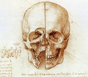
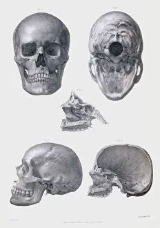
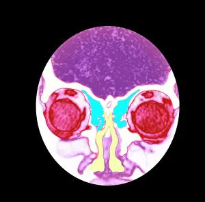
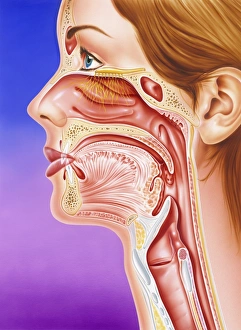
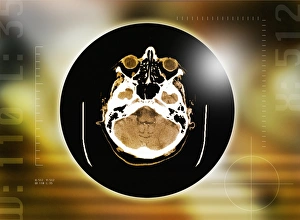
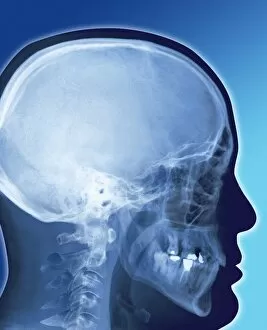
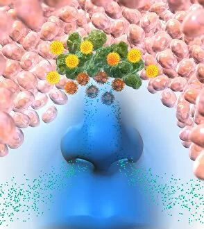
"Exploring the Intricacies of Sinuses: Unveiling the Hidden Marvels within Our Skull" Step into the realm of anatomical exploration as we delve deep into the captivating world of sinuses. Just like Leonardo da Vinci's meticulous study of skull anatomy, let us embark on a journey to unravel the mysteries surrounding these fascinating cavities. Through CT scans and sectional dissected heads from different eras, we witness how sinuses intricately intertwine with our nasal passages, humerus, vertebrae, and even our brain. The 19th-century artwork showcases their presence in stunning detail, reminding us that these hidden structures have captivated curious minds for centuries. As we peer into brain sinuses depicted in mesmerizing 1825 artwork, we gain a newfound appreciation for their vital role in maintaining cerebral health. These intricate networks of veins not only ensure proper blood flow but also contribute to overall brain function. Intriguingly enough, lymph nodes are also intertwined with sinus pathways. Artwork from C013 / 4632 and C013 / 4631 highlights their presence within this complex system – an essential component ensuring immune responses remain efficient and effective. The connection between nose, mouth, throat becomes evident as we explore further, and is through these interconnected passageways that air flows seamlessly while allowing us to taste flavors or savor fragrances. Sinuses play a crucial role here too by regulating airflow and filtering out impurities before they reach our lungs. CT scans provide valuable insights into sinus structure variations among individuals – shedding light on why some may experience chronic congestion or infections more frequently than others. Understanding these differences can aid medical professionals in providing personalized care tailored to each patient's unique needs. So let us marvel at the wonders concealed within our skulls – where art meets science; where Leonardo da Vinci's passion for anatomy merges with modern technology; where ancient knowledge blends seamlessly with contemporary discoveries.