
Sem Collection
"Exploring the Microcosmos: Unveiling the Hidden World with SEM" Step into a world unseen by the naked eye


All Professionally Made to Order for Quick Shipping
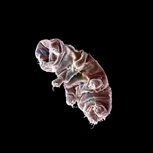
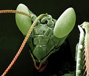
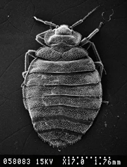
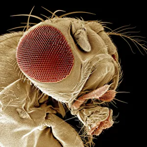
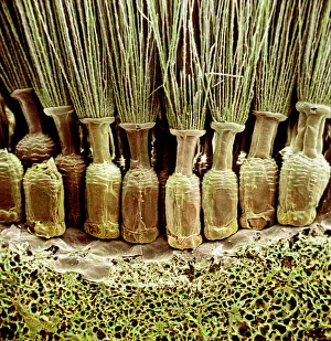
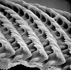
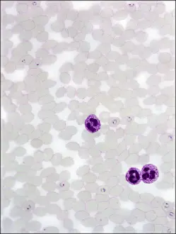
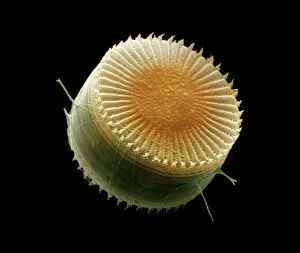
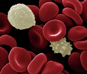
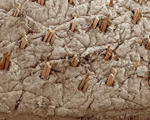
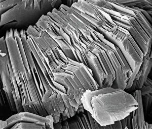
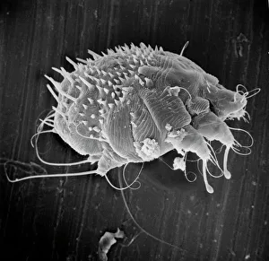
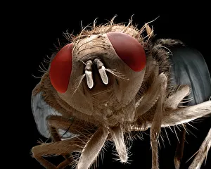
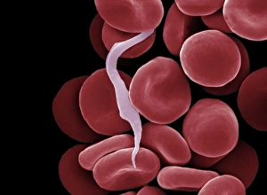
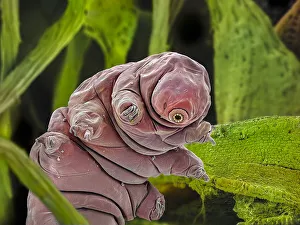
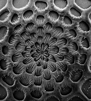
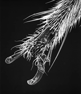
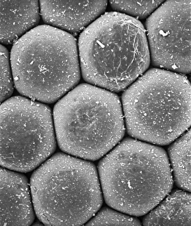
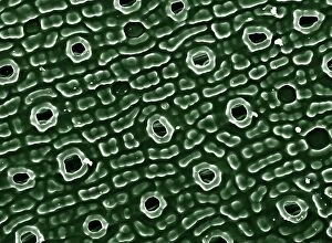
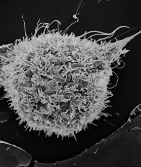
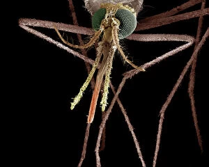
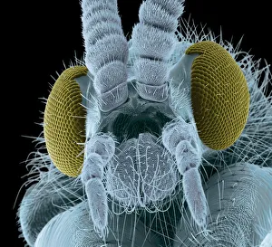
"Exploring the Microcosmos: Unveiling the Hidden World with SEM" Step into a world unseen by the naked eye, where tiny creatures and structures come to life under the powerful lens of a Scanning Electron Micrograph (SEM). In this captivating journey, we encounter fascinating subjects that range from insects to minerals, each revealing intricate details that astound. First up is the Praying Mantis, captured at a magnification of x30. The SEM exposes its delicate features in stunning clarity, showcasing nature's remarkable design. Moving on to another creature, we delve deeper into the microscopic realm with a Tardigrade or 'Water Bear' at an astonishing x1250 magnification. This resilient organism amazes us with its ability to survive extreme conditions. Shifting our focus towards minerals, Crysotile asbestos takes center stage. Despite its controversial reputation due to health risks associated with exposure, this SEM image highlights its unique fibrous structure. Santos-Dumont/Sem introduces us to aviation history as we witness intricate mechanical components through electron microscopy. The liver comes next—a vital organ responsible for numerous bodily functions—revealing its complex cellular architecture when examined under an SEM lens. Bed bugs also make their appearance; Cimex lectularius showcases their minuscule yet menacing presence in our homes. Continuing our exploration of insect lifeforms, we encounter a Fruit Fly at x300 magnification—an exquisite display of wings and sensory organs that aid these tiny creatures in survival. Taraxacum officinale or dandelion fruiting head adds vibrant beauty amidst scientific marvels. Delving further into nature's wonders brings us snail teeth—an unexpected revelation of intricately patterned dental structures designed for devouring vegetation. Plasmodium sp. , known as malarial parasite follows suit—a haunting reminder of both danger and scientific progress towards combating infectious diseases.