
Scientific Imaging Collection
Scientific Imaging: Unveiling the Inner Secrets of the Human Body In the realm of scientific exploration


All Professionally Made to Order for Quick Shipping
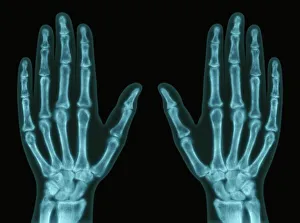
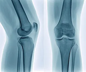
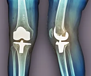
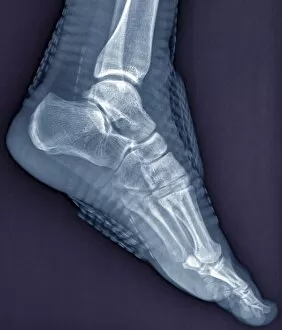
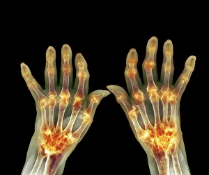
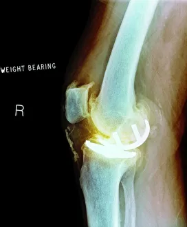
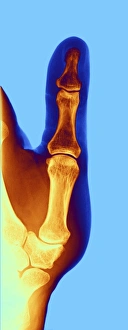
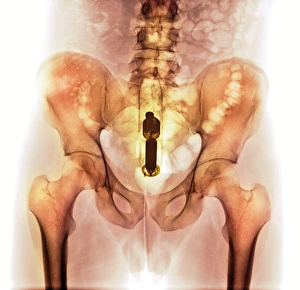
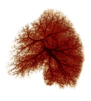
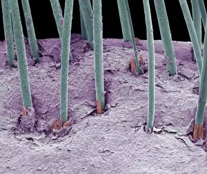
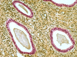
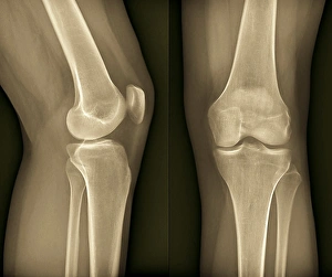
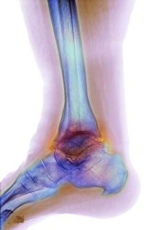
Scientific Imaging: Unveiling the Inner Secrets of the Human Body In the realm of scientific exploration, imaging techniques have revolutionized our understanding of the intricate workings within our bodies. With hands and X-rays as their tools, scientists delve into a world unseen by naked eyes. A healthy knee reveals its hidden marvels through an X-ray, showcasing its robust structure that supports our every move. On the other hand, total knee replacement X-rays demonstrate how medical advancements can restore mobility and alleviate pain. Similar to knees, a healthy ankle joint captured in an X-ray unveils its strength and flexibility. However, when scoliosis affects the spine, X-rays expose its abnormal curvature, prompting further investigation for treatment options. The power extends beyond bones; it delves into diseases like rheumatoid arthritis that leave their mark on joints. Through detailed X-rays, doctors gain insights into disease progression and tailor treatments accordingly. Beyond skeletal structures lie vital organs such as lungs—X-rays provide invaluable information about their health status. These images aid in diagnosing conditions ranging from pneumonia to lung cancer at early stages when intervention is most effective. Osteoarthritis plagues many with knee pain; however, through meticulous examination using X-rays, physicians identify characteristic signs like joint space narrowing or bone spurs—a roadmap guiding personalized care plans. Moving away from traditional radiography techniques lies scanning electron microscopy (SEM), which unlocks microscopic wonders such as white blood cells—the guardians defending us against infections. SEM captures these warriors in stunning detail revealing their unique shapes and functions. Scientific imaging encompasses more than just medical applications—it embraces innovation too. The marriage between digital SLR cameras and colored X-ray technology has birthed mesmerizing visuals that blend artistry with science—an awe-inspiring fusion. Even seemingly mundane ailments like arthrosis of the hand find themselves under scrutiny through specialized hand-focused X-ray examinations. These images enable accurate diagnosis and guide hand surgeons in restoring dexterity.