
Scapula Collection
The scapula, also known as the shoulder blade, is a crucial bone in our body that plays a significant role in our mobility and stability


All Professionally Made to Order for Quick Shipping
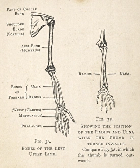
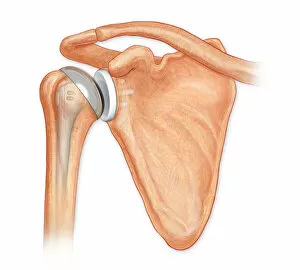
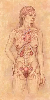
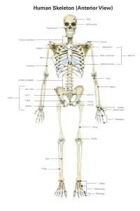
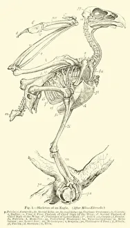
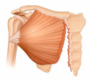
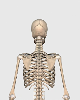
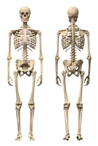
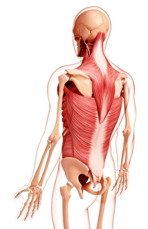
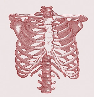
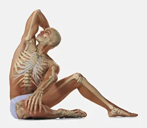
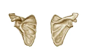
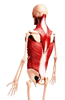
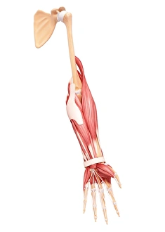
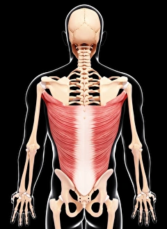
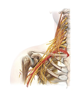
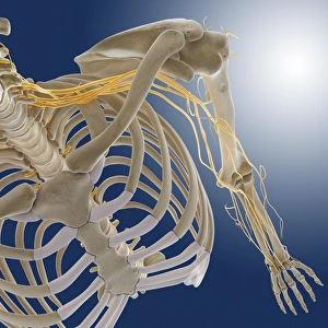
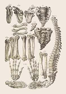
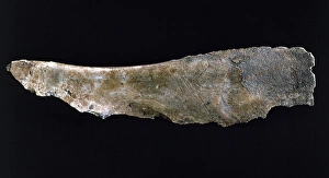
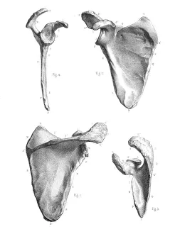
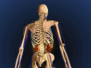
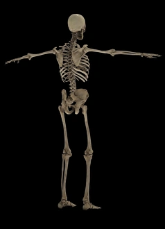
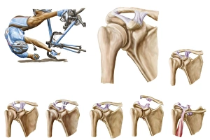
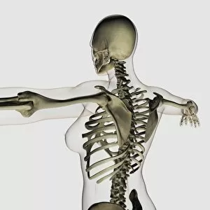
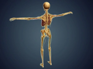
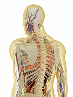
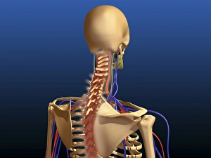
The scapula, also known as the shoulder blade, is a crucial bone in our body that plays a significant role in our mobility and stability. It forms part of the pectoral girdle and connects the upper arm to the thorax. When we examine diagrams of the bones of the hand and arm, we can see how intricately connected they are to the scapula. This connection allows us to perform various movements with precision and control. In an anterior view total shoulder joint repair image, we witness medical advancements aimed at restoring functionality to this vital joint. The intricate procedure highlights just how important it is to maintain a healthy scapula for optimal movement. Shoulder muscles artwork showcases their attachment points on the scapula, emphasizing their role in providing strength and stability during physical activities. Understanding these muscles helps us appreciate their contribution to everyday tasks like lifting or throwing. A front view of female anatomy highlighting the endocrine system reminds us that even though not directly related to the scapula, every part of our body works together harmoniously. Hormones secreted by glands within this system influence bone health and development. An anterior view of human skeletal system with labels gives us a comprehensive understanding of where exactly our scapula fits into this complex framework. It serves as an anchor point for numerous ligaments and tendons essential for proper functioning. Pictograms on an ox scapula depict ancient rituals performed to ward off danger—a testament to how cultures throughout history recognized its significance beyond mere anatomy. These artifacts remind us that humans have long understood its importance in daily life. The skeleton of an eagle after Milne-Edwards engraving demonstrates nature's adaptation at its finest—the bird's wingspan relies heavily on strong shoulder blades (scapulas) allowing it effortless flight through vast skies. Haydon's Curtius engraving captures another artistic representation showcasing human form—this time focusing on muscular structure including the scapula.