
Scanning Electron Microscope Image Collection (page 5)
"Unlocking the Hidden World: Exploring Fascinating Microcosms through Scanning Electron Microscope Images" Delving into the intricate realms of nature and science


All Professionally Made to Order for Quick Shipping
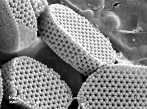
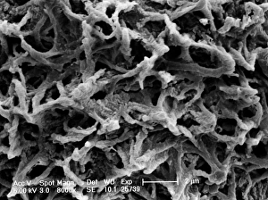
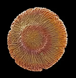
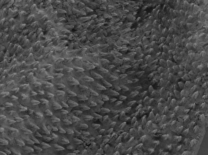
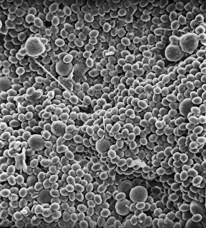
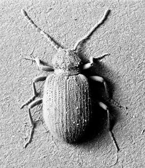
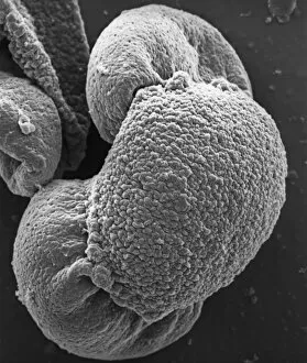
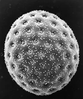
"Unlocking the Hidden World: Exploring Fascinating Microcosms through Scanning Electron Microscope Images" Delving into the intricate realms of nature and science, scanning electron microscope (SEM) images offer a mesmerizing glimpse into the microscopic wonders that surround us. From Crysotile asbestos fibers to the complex structure of a liver cell, these captivating images reveal an unseen universe. Witness the astonishing detail of a Cimex lectularius, commonly known as a bed bug, showcasing its exoskeleton with remarkable precision. Zoom in further to observe Taraxacum officinale's fruiting head, where each tiny seed is meticulously arranged like nature's own masterpiece. Marvel at snail teeth magnified to unprecedented levels, exposing their unique formation designed for efficient feeding. Observe Plasmodium sp. , the malarial parasite responsible for causing one of humanity's deadliest diseases - its intricate morphology unraveled before your eyes. Dive deeper into geological wonders as Kaolinite crystals come alive under SEM imaging; their delicate structures resembling miniature works of art. Explore Sarcoptes scabiei, better known as scabies mites, revealing their minuscule bodies adapted for survival within human skin crevices. Venture into fungal realms with Aspergillus species captured in stunning detail; witness their filamentous hyphae intertwining intricately like an otherworldly web. Discover new life emerging from caterpillar eggs under high-resolution microscopy - each egg harboring potential transformation and growth. Uncover secrets hidden within blackfly antennae; marvel at Anopheles gambiae mosquitoes' delicate features that enable them to transmit deadly diseases such as malaria. These SEM images provide invaluable insights into our world's smallest inhabitants and contribute to scientific advancements aimed at understanding and combating various threats they pose. Through scanning electron microscope imagery, we are transported beyond what meets the naked eye – unlocking mysteries that shape our understanding of the natural world.