Fraud Blocker Skip to main content

Salivary Gland Collection

"Exploring the Marvels of Salivary Glands

Background imageSalivary Gland Collection: Diagram showing inside of mouth and salivary glands

Diagram showing inside of mouth and salivary glands

Background imageSalivary Gland Collection: Salivary gland stone, X-ray

Salivary gland stone, X-ray
Salivary gland stone. X-ray of the jaw of a 36-year-old patient with a stone (white, lower left) in the right submaxillary gland

Background imageSalivary Gland Collection: Eastern equine encephalitis virus, TEM

Eastern equine encephalitis virus, TEM
Eastern equine encephalitis virus. Coloured transmission electron micrograph (TEM) of eastern equine encephalitis (EEE) virus particles (Alphavirus sp. orange) in the salivary gland of a mosquito

Background imageSalivary Gland Collection: Woman sat with leg outstretched to one side, other leg folded in, arm behind head, skeleton

Woman sat with leg outstretched to one side, other leg folded in, arm behind head, skeleton, lymphatic system illustration overlay

Background imageSalivary Gland Collection: St. Louis encephalitis virus particles C016 / 9453

St. Louis encephalitis virus particles C016 / 9453
St. Louis encephalitis virus particles. Coloured transmission electron micrograph (TEM) of a section through a mosquito salivary gland containing numerous Saint Louis encephalitis virus particles

Background imageSalivary Gland Collection: Chromosomes, light micrograph C016 / 6354

Chromosomes, light micrograph C016 / 6354
Chromosomes, light micrograph. This micrograph was obtained in 1934 during studies of chromosome structure in the salivary glands of Sciara (a genus of fungus gnats)

Background imageSalivary Gland Collection: Blood clot, SEM C014 / 0381

Blood clot, SEM C014 / 0381
Blood clot. Coloured scanning electron micrograph (SEM) of a blood clot (thrombus) in an arteriole (small blood vessel) of a salivary gland

Background imageSalivary Gland Collection: Blood clot, SEM C014 / 0380

Blood clot, SEM C014 / 0380
Blood clot. Coloured scanning electron micrograph (SEM) of a blood clot (thrombus) in an arteriole (small blood vessel) of a salivary gland

Background imageSalivary Gland Collection: Salivary gland anatomy, artwork

Salivary gland anatomy, artwork
Salivary gland anatomy. Artwork of a sectioned salivary gland, showing its acinar structure where salivary fluids are secreted in lobes (three shown)

Background imageSalivary Gland Collection: Enlarged 60 times. Line engraving, French, 19th century

Enlarged 60 times. Line engraving, French, 19th century
HUMAN SALIVARY GLAND. Enlarged 60 times. Line engraving, French, 19th century

Background imageSalivary Gland Collection: Salivary gland, light micrograph

Salivary gland, light micrograph
Salivary gland. Coloured light micrograph of a section through a sublingual salivary gland, which is situated directly under the tongue

Background imageSalivary Gland Collection: Artwork showing lymph nodes in the neck

Artwork showing lymph nodes in the neck
Lymph nodes. Artwork showing the position of lymph nodes (green) in the neck. Lymph nodes are part of the lymphatic system, which helps protect against diseases and infections

Background imageSalivary Gland Collection: Parotid salivary gland, light micrograph

Parotid salivary gland, light micrograph
Parotid salivary gland. Light micrograph of a section through a parotid salivary gland showing serous acini (green) and their ducts (bright green)

Background imageSalivary Gland Collection: Giant chromosomes, light micrograph

Giant chromosomes, light micrograph. These giant (polytene) chromosomes are from the salivary glands of the fruit fly Drosophila melanogaster

Background imageSalivary Gland Collection: Giant chromosome, light micrograph

Giant chromosome, light micrograph

Background imageSalivary Gland Collection: Aquaporin roles in the body

Aquaporin roles in the body. Aquaporins are membrane proteins that form channels (lower right) that help water molecules pass in and out of cells


All Professionally Made to Order for Quick Shipping

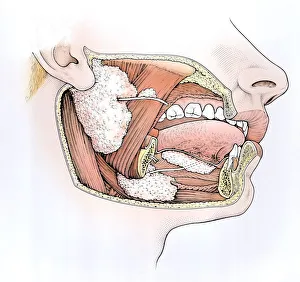
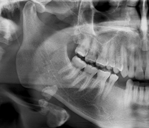
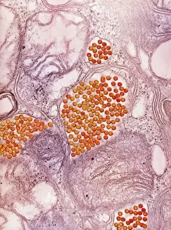
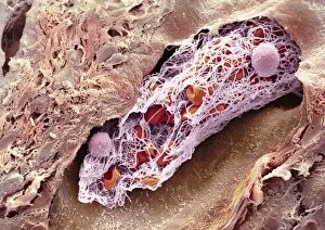
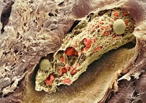
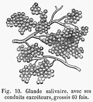
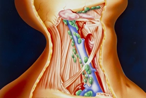
"Exploring the Marvels of Salivary Glands: A Gateway to Digestion and Disease" Delve into the intricate world of salivary glands with this cross-section biomedical illustration, showcasing their vital role in our digestive system. Witness the inner workings of your mouth as this diagram reveals the location and function of salivary glands, essential for saliva production. Uncover a common challenge faced by some individuals - a salivary gland stone - through an X-ray image that highlights its presence and potential complications. Discover how even viruses can target these important glands, such as Eastern equine encephalitis virus, as revealed by a TEM image capturing their interaction at a microscopic level. Immerse yourself in an intriguing composition featuring a woman's relaxed pose overlaid with illustrations depicting her skeleton and lymphatic system – emphasizing the interconnectedness between our bodies' systems, including salivary glands. Gain insights into St. Louis encephalitis virus particles through captivating images C016 / 9454 and C016 / 9453, shedding light on their impact on these crucial glands. Return to exploring salivary gland stones once more with additional X-ray images that showcase different cases – highlighting both their prevalence and importance in medical diagnosis. Expand your knowledge beyond salivary glands as you encounter chromosomes captured under a light microscope (C016 / 6354) or witness a blood clot up close through SEM imaging (C014 / 0381). Join us on this fascinating journey where we unravel the mysteries surrounding our salivary glands – from aiding digestion to battling diseases – reminding us just how remarkable our bodies truly are.