Fraud Blocker Skip to main content

Rna Virus Collection (page 3)

RNA viruses are a diverse group of infectious agents that have captured the attention of scientists and researchers worldwide

Background imageRna Virus Collection: Enterovirus particle C014 / 4898

Enterovirus particle C014 / 4898
Enterovirus particle. Computer artwork of an enterovirus particle (virion), showing the structure of the capsid (outer shell)

Background imageRna Virus Collection: Human poliovirus, molecular model

Human poliovirus, molecular model
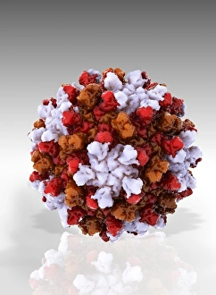
Human poliovirus particle. Computer model of the capsid of the human poliovirus. The capsid is a protein coat that encloses the viruss genetic information (genome), stored as RNA (ribonucleic acid)

Background imageRna Virus Collection: Enterovirus particles C014 / 4899

Enterovirus particles C014 / 4899
Enterovirus particles. Computer artwork of enterovirus particles (virion). Enteroviruses are a genus of non-enveloped positive-sense single-stranded RNA viruses associated with several human

Background imageRna Virus Collection: HIV particles, artwork C016 / 8659

HIV particles, artwork C016 / 8659
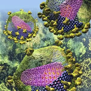
HIV particles, computer artwork. HIV (human immunodeficiency virus) causes AIDS (acquired immune deficiency syndrome). The virus consists of an RNA (ribonucleic acid) genome (pink)

Background imageRna Virus Collection: HIV particles, artwork C016 / 8658

HIV particles, artwork C016 / 8658
HIV particles, computer artwork. HIV (human immunodeficiency virus) causes AIDS (acquired immune deficiency syndrome). The virus consists of an RNA (ribonucleic acid) genome (pink)

Background imageRna Virus Collection: HIV particles, artwork C016 / 8655

HIV particles, artwork C016 / 8655
HIV particles, computer artwork. HIV (human immunodeficiency virus) causes AIDS (acquired immune deficiency syndrome). The virus consists of an RNA (ribonucleic acid) genome (pink)

Background imageRna Virus Collection: HIV particles, artwork C016 / 8654

HIV particles, artwork C016 / 8654
HIV particles, computer artwork. HIV (human immunodeficiency virus) causes AIDS (acquired immune deficiency syndrome). The virus consists of an RNA (ribonucleic acid) genome (pink)

Background imageRna Virus Collection: HIV particles, artwork C016 / 8652

HIV particles, artwork C016 / 8652
HIV particles, computer artwork. HIV (human immunodeficiency virus) causes AIDS (acquired immune deficiency syndrome). The virus consists of an RNA (ribonucleic acid) genome (pink)

Background imageRna Virus Collection: HIV particles, artwork C016 / 8653

HIV particles, artwork C016 / 8653
HIV particles, computer artwork. HIV (human immunodeficiency virus) causes AIDS (acquired immune deficiency syndrome). The virus consists of an RNA (ribonucleic acid) genome (pink)

Background imageRna Virus Collection: HIV particle, artwork C016 / 8649

HIV particle, artwork C016 / 8649
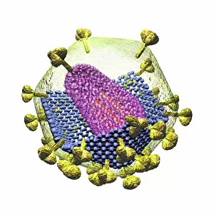
HIV particle, computer artwork. HIV (human immunodeficiency virus) causes AIDS (acquired immune deficiency syndrome). The virus consists of an RNA (ribonucleic acid) genome

Background imageRna Virus Collection: HIV particle, artwork C016 / 8651

HIV particle, artwork C016 / 8651
HIV particle, computer artwork. HIV (human immunodeficiency virus) causes AIDS (acquired immune deficiency syndrome). The virus consists of an RNA (ribonucleic acid) genome

Background imageRna Virus Collection: HIV particle, artwork C016 / 8650

HIV particle, artwork C016 / 8650
HIV particle, computer artwork. HIV (human immunodeficiency virus) causes AIDS (acquired immune deficiency syndrome). The virus consists of an RNA (ribonucleic acid) genome

Background imageRna Virus Collection: HIV particle, artwork C016 / 8648

HIV particle, artwork C016 / 8648
HIV particle, computer artwork. HIV (human immunodeficiency virus) causes AIDS (acquired immune deficiency syndrome). The virus consists of an RNA (ribonucleic acid) genome (pink)

Background imageRna Virus Collection: HIV particle, artwork C016 / 8647

HIV particle, artwork C016 / 8647
HIV particle, computer artwork. HIV (human immunodeficiency virus) causes AIDS (acquired immune deficiency syndrome). The virus consists of an RNA (ribonucleic acid) genome (pink)

Background imageRna Virus Collection: HIV particle, artwork C016 / 8646

HIV particle, artwork C016 / 8646
HIV particle, computer artwork. HIV (human immunodeficiency virus) causes AIDS (acquired immune deficiency syndrome). The virus consists of an RNA (ribonucleic acid) genome (pink)

Background imageRna Virus Collection: Influenza virus, artwork C016 / 8349

Influenza virus, artwork C016 / 8349
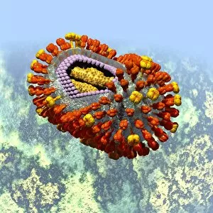
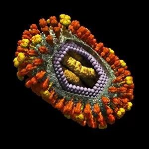
Influenza virus. Cut-away computer artwork of an influenza (flu) virus particle (virion). In each particles lipid envelope (green) are two types of protein spike, haemagglutinin (H)

Background imageRna Virus Collection: Influenza virus, artwork C016 / 8348

Influenza virus, artwork C016 / 8348
Influenza virus. Cut-away computer artwork of an influenza (flu) virus particle (virion). In each particles lipid envelope (green) are two types of protein spike, haemagglutinin (H)

Background imageRna Virus Collection: Influenza virus, artwork C016 / 8347

Influenza virus, artwork C016 / 8347
Influenza virus. Cut-away computer artwork of an influenza (flu) virus particle (virion). In each particles lipid envelope (green) are two types of protein spike, haemagglutinin (H)

Background imageRna Virus Collection: Influenza virus, artwork C016 / 8346

Influenza virus, artwork C016 / 8346
Influenza virus. Cut-away computer artwork of an influenza (flu) virus particle (virion). In each particles lipid envelope (green) are two types of protein spike, haemagglutinin (H)

Background imageRna Virus Collection: Influenza virus, artwork C016 / 8344

Influenza virus, artwork C016 / 8344
Influenza virus. Cut-away computer artwork of an influenza (flu) virus particle (virion). In each particles lipid envelope (green) are two types of protein spike, haemagglutinin (H)

Background imageRna Virus Collection: Influenza virus, artwork C016 / 8345

Influenza virus, artwork C016 / 8345
This image may not be used in educational posters Influenza virus. Cut-away computer artwork of an influenza (flu) virus particle (virion)

Background imageRna Virus Collection: Influenza virus, artwork C016 / 8342

Influenza virus, artwork C016 / 8342
Influenza virus. Computer artwork of an influenza (flu) virus particle (virion). In each particles lipid envelope (green) are two types of protein spike, haemagglutinin (H, red) and neuraminidase (N)

Background imageRna Virus Collection: Influenza virus, artwork C016 / 8341

Influenza virus, artwork C016 / 8341
Influenza virus. Computer artwork of an influenza (flu) virus particle (virion). In each particles lipid envelope (green) are two types of protein spike, haemagglutinin (H, red) and neuraminidase (N)

Background imageRna Virus Collection: Influenza virus, artwork C016 / 8340

Influenza virus, artwork C016 / 8340
Influenza virus. Computer artwork of an influenza (flu) virus particle (virion). In each particles lipid envelope (green) are two types of protein spike, haemagglutinin (H, red) and neuraminidase (N)

Background imageRna Virus Collection: Tomato bushy stunt virus, molecular model

Tomato bushy stunt virus, molecular model
Tomato bushy stunt virus. Computer model showing the molecular structure of a tomato bushy stunt virus (TBSV) capsid (outer protein coat)

Background imageRna Virus Collection: Cell infected with HIV, SEM C014 / 0581

Cell infected with HIV, SEM C014 / 0581
Cell infected with HIV. Coloured scanning electron micrograph (SEM) of HIV particles (round) budding from the membrane of a host cell

Background imageRna Virus Collection: Cell infected with HIV, SEM C014 / 0580

Cell infected with HIV, SEM C014 / 0580
Cell infected with HIV. Coloured scanning electron micrograph (SEM) of HIV particles (round) budding from the membrane of a host cell

Background imageRna Virus Collection: Cell infected with HIV, SEM C014 / 0579

Cell infected with HIV, SEM C014 / 0579
Cell infected with HIV. Coloured scanning electron micrograph (SEM) of HIV particles (round) budding from the membrane of a host cell

Background imageRna Virus Collection: Cell infected with HIV, SEM C017 / 8338

Cell infected with HIV, SEM C017 / 8338
Cell infected with HIV. Coloured scanning electron micrograph (SEM) of HIV particles (orange) budding from the membrane of a host cell

Background imageRna Virus Collection: Cell infected with HIV, SEM C017 / 8339

Cell infected with HIV, SEM C017 / 8339
Cell infected with HIV. Coloured scanning electron micrograph (SEM) of HIV particles (orange) budding from the membrane of a host cell

Background imageRna Virus Collection: Cell infected with HIV, SEM C017 / 8337

Cell infected with HIV, SEM C017 / 8337
Cell infected with HIV. Coloured scanning electron micrograph (SEM) of HIV particles (yellow) budding from the membrane of a host cell

Background imageRna Virus Collection: Cell infected with HIV, SEM C017 / 8336

Cell infected with HIV, SEM C017 / 8336
Cell infected with HIV. Coloured scanning electron micrograph (SEM) of HIV particles (pink) budding from the membrane of a host cell

Background imageRna Virus Collection: Mouse leukaemia virus and T-cell, SEM C017 / 8308

Mouse leukaemia virus and T-cell, SEM C017 / 8308
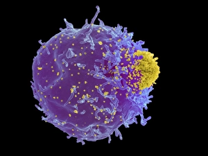
Mouse leukaemia virus. Coloured scanning electron micrograph (SEM) of mouse leukaemia virus particles (yellow) budding out of a T-lymphocyte (or T-cell, purple)

Background imageRna Virus Collection: Budding HIV particles, SEM C017 / 8302

Budding HIV particles, SEM C017 / 8302
Budding HIV particles. Coloured scanning electron micrograph (SEM) of human immunodeficiency virus (HIV) particles (yellow) budding from the surface of a T lymphocyte from the H9 cell line

Background imageRna Virus Collection: Influenza virus, artwork C018 / 2894

Influenza virus, artwork C018 / 2894
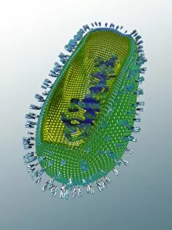
Influenza virus. Cut-away computer artwork of an influenza (flu) virus particle (virion). In each particles lipid envelope (blue) are two types of protein spike

Background imageRna Virus Collection: HIV particle, artwork C018 / 2898

HIV particle, artwork C018 / 2898
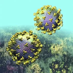
HIV particle, computer artwork. HIV (human immunodeficiency virus) causes AIDS (acquired immune deficiency syndrome). The virus consists of an RNA (ribonucleic acid) genome

Background imageRna Virus Collection: HIV particle, artwork C018 / 2897

HIV particle, artwork C018 / 2897
HIV particle, computer artwork. HIV (human immunodeficiency virus) causes AIDS (acquired immune deficiency syndrome). The virus consists of an RNA (ribonucleic acid) genome

Background imageRna Virus Collection: HIV particle, artwork C018 / 2899

HIV particle, artwork C018 / 2899
HIV particle, computer artwork. HIV (human immunodeficiency virus) causes AIDS (acquired immune deficiency syndrome). The virus consists of an RNA (ribonucleic acid) genome

Background imageRna Virus Collection: Influenza virus, artwork C018 / 2893

Influenza virus, artwork C018 / 2893
Influenza virus. Cut-away computer artwork of an influenza (flu) virus particle (virion). In each particles lipid envelope (blue) are two types of protein spike

Background imageRna Virus Collection: Influenza virus, artwork C018 / 2891

Influenza virus, artwork C018 / 2891
Influenza virus. Cut-away computer artwork of an influenza (flu) virus particle (virion). In each particles lipid envelope (blue) are two types of protein spike

Background imageRna Virus Collection: HIV particle, artwork C018 / 2895

HIV particle, artwork C018 / 2895
HIV particle, computer artwork. HIV (human immunodeficiency virus) causes AIDS (acquired immune deficiency syndrome). The virus consists of an RNA (ribonucleic acid) genome

Background imageRna Virus Collection: HIV particle, artwork C018 / 2900

HIV particle, artwork C018 / 2900
HIV particle, computer artwork. HIV (human immunodeficiency virus) causes AIDS (acquired immune deficiency syndrome). The virus consists of an RNA (ribonucleic acid) genome

Background imageRna Virus Collection: HIV particle, artwork C018 / 2896

HIV particle, artwork C018 / 2896
HIV particle, computer artwork. HIV (human immunodeficiency virus) causes AIDS (acquired immune deficiency syndrome). The virus consists of an RNA (ribonucleic acid) genome

Background imageRna Virus Collection: Influenza virus, artwork C018 / 2890

Influenza virus, artwork C018 / 2890
Influenza virus. Cut-away computer artwork of an influenza (flu) virus particle (virion). In each particles lipid envelope (green) are two types of protein spike

Background imageRna Virus Collection: Influenza virus, artwork C018 / 2892

Influenza virus, artwork C018 / 2892
Influenza virus. Cut-away computer artwork of an influenza (flu) virus particle (virion). In each particles lipid envelope (brown) are two types of protein spike

Background imageRna Virus Collection: Avian influenza virus, TEM C016 / 5843

Avian influenza virus, TEM C016 / 5843
Avian influenza virus, type A strain H7N9, coloured transmission electron micrograph (TEM). This virus first emerged in the human population in China, in March 2013

Background imageRna Virus Collection: Avian influenza virus, TEM C016 / 5841

Avian influenza virus, TEM C016 / 5841
Avian influenza virus, type A strain H7N9, coloured transmission electron micrograph (TEM). This virus first emerged in the human population in China, in March 2013

Background imageRna Virus Collection: Avian influenza virus, TEM C016 / 5842

Avian influenza virus, TEM C016 / 5842
Avian influenza virus, type A strain H7N9, coloured transmission electron micrograph (TEM). This virus first emerged in the human population in China, in March 2013


All Professionally Made to Order for Quick Shipping

RNA viruses are a diverse group of infectious agents that have captured the attention of scientists and researchers worldwide. From the intricate structure of HIV particles to the menacing Norovirus particles observed under a transmission electron microscope (TEM), these tiny entities hold immense significance in our understanding of viral diseases. Influenza, one of the most well-known RNA viruses, has been depicted through computer artwork, showcasing its complex architecture and highlighting its ability to mutate rapidly. TEM images further reveal the true face of influenza virus particles, resembling miniature spheres with spikes protruding from their surface. Hepatitis C viruses also fall into this category, as revealed by TEM imaging. These minute structures can wreak havoc on liver cells and cause chronic infections if left untreated. The artistic representation of rotavirus particle emphasizes its distinctive wheel-like shape, which is responsible for causing severe diarrhea in young children. Molecular models depicting rhinovirus alongside antibodies shed light on potential therapeutic strategies against common colds caused by this RNA virus. Similarly, an SEM image showcases an HIV-infected macrophage - a key player in spreading this deadly virus throughout the body. The captivating artwork capturing infectious bronchitis virus (IBV) highlights how RNA viruses can affect not only humans but also animals like poultry. L-A virus and Coxsackievirus are two more examples that demonstrate the wide range of hosts susceptible to RNA viral infections. As we delve deeper into studying these enigmatic entities known as RNA viruses, we uncover their remarkable adaptability and ability to exploit host cells for replication. Understanding their mechanisms will undoubtedly pave the way for novel treatments and preventive measures against these relentless adversaries that continue to challenge human health globally.