
Research Subjects Collection
"Exploring the Microscopic World: Fascinating Research Subjects Unveiled" Delving into the realm of scientific research


All Professionally Made to Order for Quick Shipping
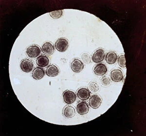
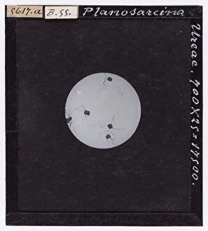
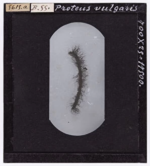
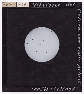
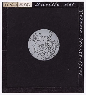
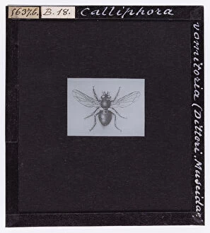
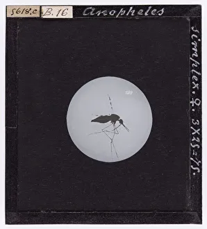
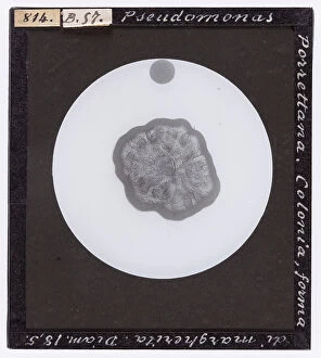
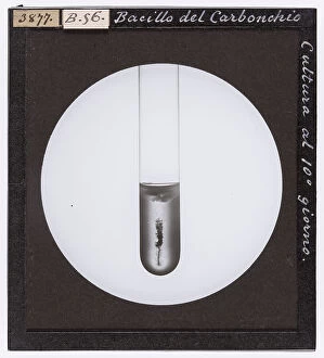
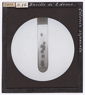
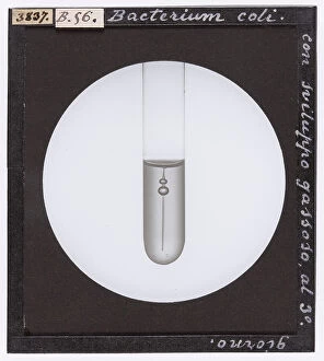
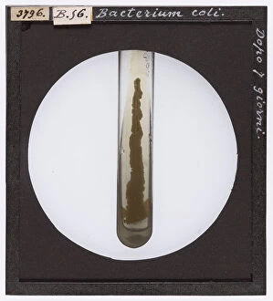
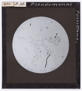
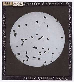
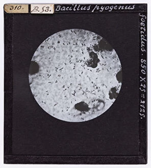
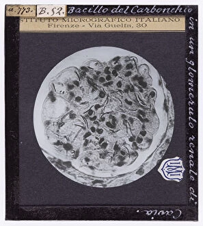
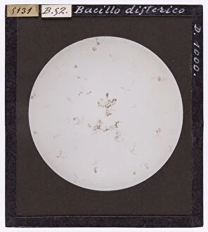
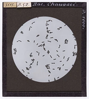
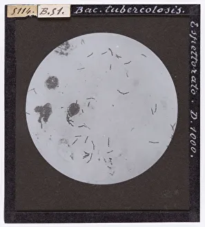
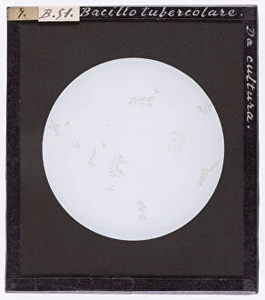
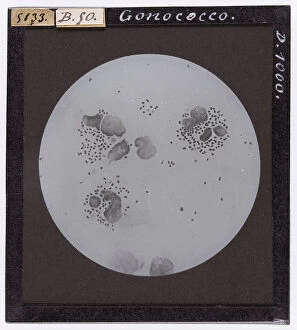
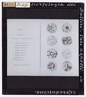
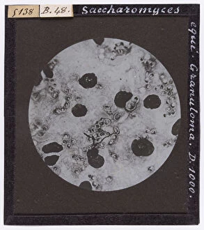
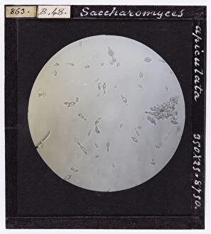
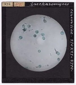
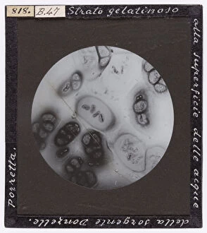
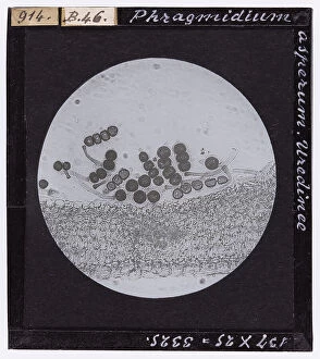
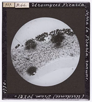
"Exploring the Microscopic World: Fascinating Research Subjects Unveiled" Delving into the realm of scientific research, we uncover a captivating world that lies beyond our naked eye. Planoarcina Ureae, enlarged under a microscope, reveals intricate patterns and structures that astound the mind. Similarly, the microscopic enlargement of 'Proteus Vulgaris' showcases its unique characteristics and offers valuable insights. As we journey further into this hidden universe, we encounter the cholera vibrio magnified under a microscope. Its distinct shape and features provide scientists with crucial information in combating this deadly disease. The bacillus also takes center stage as it is examined closely under microscopic lenses, unraveling secrets yet to be discovered. Intriguingly, tetanus bacillus captures our attention when observed at an enlarged scale through a microscope. The sporiferous variant adds another layer of complexity to this study – an enigma waiting to be unraveled by dedicated researchers. The exploration continues with bacteria such as Streptococcus coming into focus under the lens. Their vibrant colors and diverse forms remind us of their omnipresence in our daily lives while urging us to understand them better for improved health outcomes. Venturing beyond medical subjects, Calliphora Vomitoria from A. Celli's "Manuale dell'igienista" transports us back in time to 1912 Torino where entomological studies were conducted meticulously. Sarcophaga carnaria joins this collection as well - both specimens serving as windows into historical research practices. Our expedition concludes with Anopheles mosquitoes taking center stage – females revealing their delicate features magnified under microscopes while males exhibit their own distinctive traits when similarly studied. These tiny creatures hold immense significance in understanding vector-borne diseases like malaria and contribute significantly towards public health efforts worldwide. Through these glimpses into various research subjects, we are reminded of humanity's relentless pursuit of knowledge.