Fraud Blocker Skip to main content

Reproductive Collection (page 18)

"Exploring the Intricate World of Reproduction: From Dinosaurs to Flowers and Beyond" In the ancient world

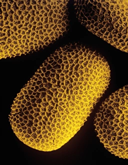
Background imageReproductive Collection: Mimosa pollen, SEM

Mimosa pollen, SEM
Mimosa pollen grains, coloured scanning electron micrograph (SEM). Magnification: x1500 when printed at 10 centimetres wide

Background imageReproductive Collection: Bay tree pollen, SEM

Bay tree pollen, SEM
Bay tree (Laurus nobilis) pollen grains, coloured scanning electron micrograph (SEM). Magnification: x1300 when printed at 10 centimetres wide

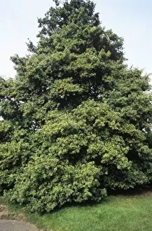
Background imageReproductive Collection: Douglas fir cones

Douglas fir cones. Branch of a Douglas fir (Pseudotsuga menziesii) trees with new growth and cones. These cones mature over the summer, producing seeds in the autumn if fertilised
Background imageReproductive Collection: Winged ant, SEM

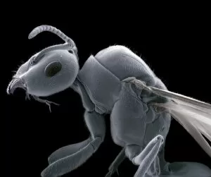
Winged ant, SEM
Winged ant, coloured scanning electron micrograph (SEM). Winged ants develop after an ant colony has been established for several years

Background imageReproductive Collection: Bee pollen basket, SEM

Bee pollen basket, SEM
Bee pollen basket, coloured scanning electron micrograph (SEM). The pollen basket (called the corbicula) is found on a bees hind legs

Background imageReproductive Collection: Hypericum pollen

Hypericum pollen. Coloured scanning electron micrograph (SEM) of pollen grains from a Hypericum sp. plant. Magnification x6000 when printed 10 centimetres wide

Background imageReproductive Collection: Orange pollen, SEM

Orange pollen, SEM
Orange (Citrus sp.) pollen grain, coloured scanning electron micrograph (SEM). Magnification: x1500 when printed at 10 centimetres wide

Background imageReproductive Collection: Mycobacterium dividing, TEM

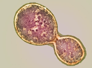
Mycobacterium dividing, TEM
Mycobacterium dividing. Coloured transmission electron micrograph (TEM) of a Mycobacterium sp. bacterium undergoing binary fission

Background imageReproductive Collection: Anthrax bacteria

Anthrax bacteria. Computer artwork of Bacillus anthracis bacteria (green) releasing spores (orange). These are Gram-positive, rod-shaped, spore-forming bacteria

Background imageReproductive Collection: Liverwort spore case, light micrograph

Liverwort spore case, light micrograph
Liverwort spore case. Light micrograph of a vertical section through a spore case (sporophyte) from a liverwort (Pellia epiphylla)

Background imageReproductive Collection: Sulphur toadstools

Sulphur toadstools. Undersides of sulphur toadstools (Tricholoma sulphureum). The gills contain million of spores, the reproductive cells of a fungus

Background imageReproductive Collection: Pine cone scale, light micrograph

Pine cone scale, light micrograph
Pine cone scale. Light micrograph of a vertical section through an ovulate scale from a Scots pine (Pinus sylvestris). This image shows the ovule (oval shape)

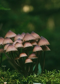
Background imageReproductive Collection: Bleeding mycena mushrooms

Bleeding mycena mushrooms (Mycena haemotopus). Bleeding mycenas are so called because they release a reddish juice when cut. They grow on rotting wood, reaching about 3-8 centimetres in height

Background imageReproductive Collection: Cryptococcus cell dividing, TEM

Cryptococcus cell dividing, TEM
Cryptococcus cell dividing. Coloured transmission electron micrograph (TEM) of the asexual budding (lower right) of a Cyptococcus sp. yeast cell

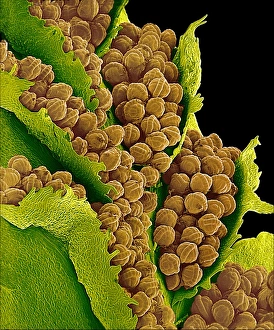
Background imageReproductive Collection: Bracken spores, SEM

Bracken spores, SEM
Bracken spores. Coloured scanning electron micrograph (SEM) of the leaves of a bracken plant (Pteridium aquilinum). The undersides of the leaves are covered in spore-containing sacs known as

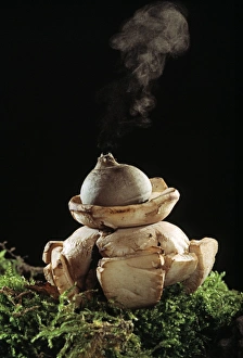
Background imageReproductive Collection: Earth star mushroom

Earth star mushroom (Geastrum triplex) growing in woodland soil. It has released (puffed) a cloud of reproductive spores (upper centre)

Background imageReproductive Collection: Horsetail spores, SEM

Horsetail spores, SEM
Horsetail spores. Coloured scanning electron micrograph (SEM) of spores of a horsetail (Equisetum sp.). The spores (orange) bear elaters (green), which expand and contract with changes in humidity

Background imageReproductive Collection: Liverwort spore cases, light micrograph

Liverwort spore cases, light micrograph
Liverwort spore cases. Light micrograph of a cross-section through five spore cases (sporophytes) from the liverwort Conocephalum conicum (Marchantiales)

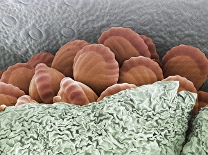
Background imageReproductive Collection: Fern spores, SEM

Fern spores, SEM
Fern spores. Coloured scanning electron micrograph (SEM) of spores (red) of a fern partly enclosed in a sporangium. The undersides of the leaves of ferns are covered in these spore-containing sacs

Background imageReproductive Collection: Atlas cedar cones

Atlas cedar cones
Atlas cedar cone (Cedrus atlantica). This is a mature, two-year-old female cone (seed)

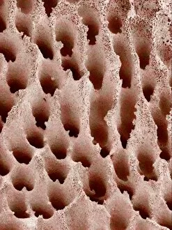
Background imageReproductive Collection: Mushroom surface, SEM

Mushroom surface, SEM
Mushroom surface. Coloured scanning electron micrograph (SEM) of the spore-bearing surface of a hen of the woods mushroom (Grifola frondosa). This surface is found on the underside of the mushroom

Background imageReproductive Collection: Calabrian pine (Pinus brutia)

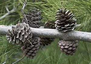
Calabrian pine (Pinus brutia)
Calabrian pine cones (Pinus brutia). Photographed in Greece, in spring

Background imageReproductive Collection: Bracken leaves, SEM

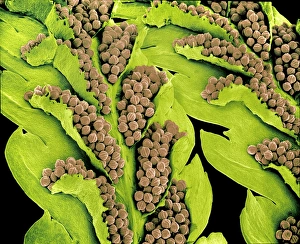
Bracken leaves, SEM
Bracken leaves. Coloured scanning electron micrograph (SEM) of the leaves of a bracken plant (Pteridium aquilinum). The undersides of the leaves are covered in spore-containing sacs known as

Background imageReproductive Collection: Mushroom gills

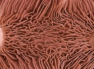
Mushroom gills. Macrophotograph of the gills of an unidentified double capped mushroom. Gills are found on the underside of the cap (fruiting body) and hold the fungal spores

Background imageReproductive Collection: Fly agaric (Amanita muscaria)

Fly agaric (Amanita muscaria) mushroom lying among ling heather (Calluna vulgaris). This fungus is poisonous if ingested, however it is often used for its hallucinogenic properties when used in small

Background imageReproductive Collection: Fungal reproduction, SEM

Fungal reproduction, SEM
Fungal reproduction. Coloured scanning electron micrograph (SEM) of the tip of a branch of a conidiophore (one type of fungal reproductive structure) with a conidia (spore) emerging from the tip

Background imageReproductive Collection: Crimson wax cap mushroom

Crimson wax cap mushroom (Hygrocybe punicea). Photographed in the New Forest, UK

Background imageReproductive Collection: White pine (Pinus strobus)

White pine (Pinus strobus) cones. Photographed in Maryland, USA

Background imageReproductive Collection: Love in the mist flower (Nigella sp. )

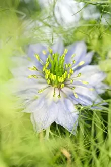
Love in the mist flower (Nigella sp. )
Love in the mist flower (Nigella sp.). The seeds of the plant, known as black cumin, are used as a spice in cookery and also medicinally as a diuretic

Background imageReproductive Collection: Primula polyanthus Hose in Hose

Primula polyanthus Hose in Hose
Polyanthus flowers (Primula polyanthus Hose in Hose ). Part of the flower has been cut away to show detail of the inside

Background imageReproductive Collection: Schlumbergera bridgesii

Schlumbergera bridgesii
Christmas cactus flowers (Schlumbergera bridgesii). This cactus is found in the Organ Mountain forests, north of Rio de Janeiro, Brazil. It is cultivated in Europe as a Christmas plant

Background imageReproductive Collection: Sessile oak (Quercus petraea)

Sessile oak (Quercus petraea)
Sessile oak tree (Quercus petraea) laden with acorns

Background imageReproductive Collection: Mixed heather

Mixed heather

Background imageReproductive Collection: Pansy flower, SEM

Pansy flower, SEM
Pansy flower reproductive parts. Coloured scanning electron micrograph (SEM) of reproductive parts from the flower of a pansy (Viola sp.) plant

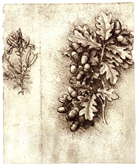
Background imageReproductive Collection: Leonardo da Vincis oak leaves and acorns

Leonardo da Vincis oak leaves and acorns
Oak leaves and acorns. Historical artwork of the leaves and fruit (acorns) of the oak tree (Quercus robur). Also seen at left are the leaves and pods of the Dyers woad plant (Isatis tinctoria)

Background imageReproductive Collection: Organic tomatoes

Organic tomatoes (Lycopersicon esculentum) ripening on a vine

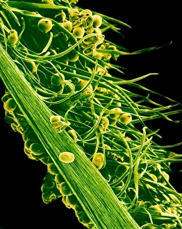
Background imageReproductive Collection: Sunflower pollination, SEM

Sunflower pollination, SEM
Sunflower pollination. Coloured scanning electron micrograph (SEM) of pollen on a stigma of a sunflower plant (Helianthus sp.)

Background imageReproductive Collection: Trifoliate orange (Poncirus trifoliata)

Trifoliate orange (Poncirus trifoliata)
Trifoliate orange fruits (Poncirus trifoliata) on their tree. This plant is closely related to Citrus, and is used as a rootstock for Citrus grafts

Background imageReproductive Collection: Field pennycress seed pod

Field pennycress seed pod
Seed pod of the field pennycress, Thlaspi arvense. The dark seeds can be seen through the seed pod. Pennycress is considered an agricultural crop weed that grows on waste ground

Background imageReproductive Collection: Sweet pea flower pollination, SEM

Sweet pea flower pollination, SEM
Sweet pea pollination. Coloured scanning electron micrograph (SEM) of pollen grains (yellow) on the stigma (green) of a sweet pea flower (Lathyrus odoratus)

Background imageReproductive Collection: Guiacum sanctum fruit

Guiacum sanctum fruit
Guiacum fruit (Guiacum sanctum). Photographed in Florida, USA

Background imageReproductive Collection: Apple (Malus Mary Charlton )

Apple (Malus Mary Charlton ) on its tree

Background imageReproductive Collection: Arum plant spadix, light micrograph

Arum plant spadix, light micrograph
Arum plant spadix. Light micrograph of a vertical section through the lower part of the spadix from a lords and ladies (Arum maculatum), or cuckoo pint, plant

Background imageReproductive Collection: Pollen grains, computer artwork

Pollen grains, computer artwork
Pollen grains. Computer artwork of pollen grains from a flower. The outer wall (exine) of each pollen grain is highly sculpted to aid the dispersal of the pollen grains

Background imageReproductive Collection: Pollen of Marsh Woundwort flower

Pollen of Marsh Woundwort flower
False colour scanning electron micrograph of pollen grains of a Marsh Woundwort. The outer walls (exine) have a netted ornamentation

Background imageReproductive Collection: Mimosa flowers, SEM

Mimosa flowers, SEM
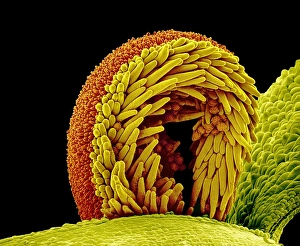
Mimosa flower stamens. Coloured scanning electron micrograph (SEM) of flowers and stamens of Mimosa sp. plants. Many stamens (male reproductive parts) are seen

Background imageReproductive Collection: Common pollen beetles feeding

Common pollen beetles feeding
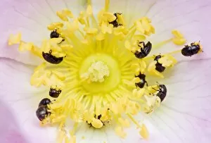
Common pollen beetles (Meligethes aeneus) feeding on pollen from a dog rose flower (Rosa canina)

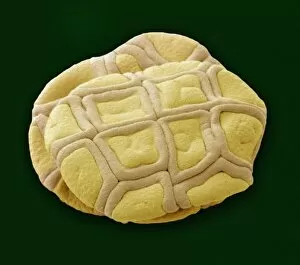
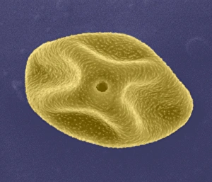
Background imageReproductive Collection: Pollen grain, SEM

Pollen grain, SEM


All Professionally Made to Order for Quick Shipping

"Exploring the Intricate World of Reproduction: From Dinosaurs to Flowers and Beyond" In the ancient world, even mighty Tyrannosaurus rex dinosaurs had their own unique ways of reproducing. Discovering fossilized evidence of their mating rituals offers a fascinating glimpse into the reproductive strategies employed by these prehistoric giants. Zooming in closer to modern times, microscopic wonders like lavender pollen grains under scanning electron microscopy (SEM) reveal intricate structures that play a crucial role in plant reproduction. Similarly, SEM images of geranium anthers and dahlia flower pollen showcase nature's attention to detail when it comes to ensuring successful pollination. Nature never ceases to amaze with its diverse range mechanisms. Take the cloudberry, for example - this Arctic fruit relies on cross-pollination facilitated by insects or wind for its continued existence. Moving beyond plants, we find ourselves exploring the realm of humans. The miracle of life unfolds within a pregnant woman's body as she nurtures new beginnings and prepares for motherhood. But reproduction is not limited to just larger organisms; even tiny creatures like honeybees have their own unique methods. SEM images capturing honeybee legs provide insight into how these industrious insects transport precious pollen from one flower to another, ensuring fertilization occurs. Venturing further into nature's hidden corners reveals captivating details about reproduction in unexpected places. Mushroom gills under SEM expose delicate structures designed for spore dispersal – a vital step in mushroom reproduction. Delving deeper still, tea flower stamens captured through SEM offer an up-close look at yet another facet of botanical fertility – showcasing intricate arrangements meant to attract pollinators and ensure successful seed production. Flowers continue enchanting us with their beauty as well as their reproductive prowess. Delphinium flowers boast vibrant colors while employing various strategies such as self-pollination or attracting specific pollinators like bees or butterflies.