Fraud Blocker Skip to main content

Protista Collection (page 4)

"Exploring the Intricate World of Protista: From Pressed Seaweed Specimens to Microscopic Wonders" In this captivating journey through the diverse realm of protista

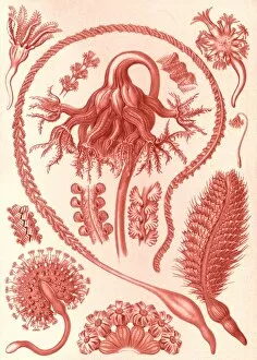
Background imageProtista Collection: Illustration shows sea pens. Pennatulida

Illustration shows sea pens. Pennatulida. - Federkorallen, 1 print : color lithograph; sheet 36 x 26 cm. 1904. Ernst Haeckel 1834 a 1919 German biologist, naturalist, philosopher, physician

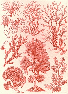
Background imageProtista Collection: Illustration shows algae. Fucoideae. - Brauntange, 1 print : lithograph; sheet 36 x 26 cm

Illustration shows algae. Fucoideae. - Brauntange, 1 print : lithograph; sheet 36 x 26 cm, 1904. Ernst Haeckel 1834 a 1919 German biologist, naturalist, philosopher, physician, professor

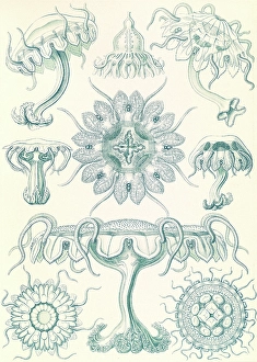
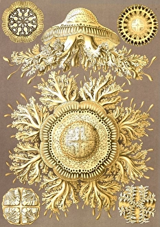
Background imageProtista Collection: Illustration shows jellyfishes. Discomedusae

Illustration shows jellyfishes. Discomedusae. - Scheibenquallen, 1 print : color lithograph; sheet 36 x 26 cm. 1904. Ernst Haeckel 1834 a 1919 German biologist, naturalist, philosopher, physician

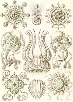
Background imageProtista Collection: Illustration shows jellyfishes. Narcomedusae

Illustration shows jellyfishes. Narcomedusae. - Spangenquallen, 1 print : color lithograph; sheet 36 x 26 cm. 1904. Ernst Haeckel 1834 a 1919 German biologist, naturalist, philosopher, physician

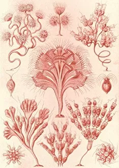
Background imageProtista Collection: Illustration shows microorganisms. Flagellata

Illustration shows microorganisms. Flagellata. - Geiklinge, 1 print : lithograph; sheet 36 x 26 cm. 1904. Ernst Haeckel 1834 a 1919 German biologist, naturalist, philosopher, physician, professor

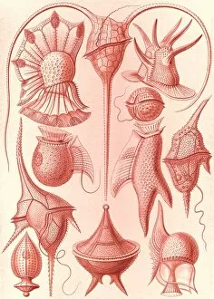
Background imageProtista Collection: Illustration shows microorganisms. Peridinea

Illustration shows microorganisms. Peridinea. - Geikelhutchen, 1 print : lithograph; sheet 36 x 26 cm. 1904. Ernst Haeckel 1834 a 1919 German biologist, naturalist, philosopher, physician, professor

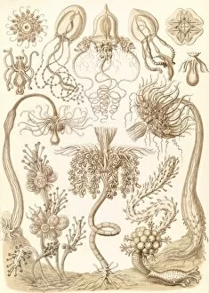
Background imageProtista Collection: Illustration shows marine invertebrates. Tubulariae

Illustration shows marine invertebrates. Tubulariae. - Rohrenpolnpen, 1 print : color lithograph; sheet 36 x 26 cm. 1904. Ernst Haeckel 1834 a 1919 German biologist, naturalist, philosopher

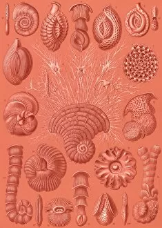
Background imageProtista Collection: Illustration shows microorganisms. Talamophora

Illustration shows microorganisms. Talamophora. - Kammerlinge, 1 print : color lithograph; sheet 36 x 26 cm. 1904. Ernst Haeckel 1834 a 1919 German biologist, naturalist, philosopher, physician

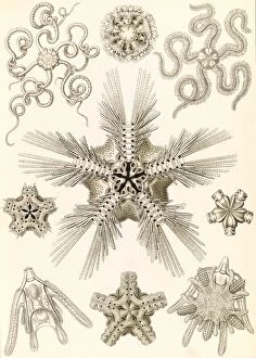
Background imageProtista Collection: Illustration shows marine invertebrates related to starfish. Ophiodea. - Schlangensterne

Illustration shows marine invertebrates related to starfish. Ophiodea. - Schlangensterne, 1 print : color lithograph; sheet 36 x 26 cm. 1904

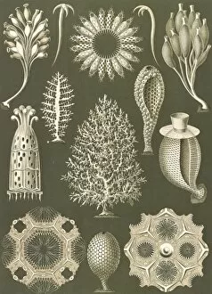
Background imageProtista Collection: Illustration shows calcareous sponges. Calcispongiae

Illustration shows calcareous sponges. Calcispongiae. - Kalkschwamme, 1 print : color lithograph; sheet 36 x 26 cm. 1904. Ernst Haeckel 1834 a 1919 German biologist, naturalist, philosopher

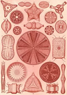
Background imageProtista Collection: Illustration shows algae. Diatomea. - Schachtellinge, 1 print : lithograph; sheet

Illustration shows algae. Diatomea. - Schachtellinge, 1 print : lithograph; sheet 36 x 26 cm. 1904. Ernst Haeckel 1834 a 1919 German biologist, naturalist, philosopher, physician, professor

Background imageProtista Collection: Illustration shows microorganisms. Ciliata

Illustration shows microorganisms. Ciliata. - Wimperlinge, 1 print : color lithograph; sheet 36 x 26 cm. 1904. Ernst Haeckel 1834 a 1919 German biologist, naturalist, philosopher, physician

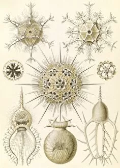
Background imageProtista Collection: Illustration shows microorganisms. Phaeodaria

Illustration shows microorganisms. Phaeodaria. - Rohrstrahlinge, 1 print : color lithograph; sheet 36 x 26 cm. 1904. Ernst Haeckel 1834 a 1919 German biologist, naturalist, philosopher, physician

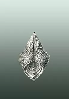
Background imageProtista Collection: Illustration showing a variety of mollusks. Acephala. - Muscheln, 1 print : lithograph

Illustration showing a variety of mollusks. Acephala. - Muscheln, 1 print : lithograph. 1904. Ernst Haeckel 1834 a 1919 German biologist, naturalist, philosopher, physician, professor

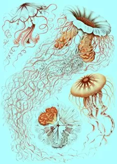
Background imageProtista Collection: Illustration showing four different types of jellyfish. Discomedusae. - Schweibenquallen

Illustration showing four different types of jellyfish. Discomedusae. - Schweibenquallen, 1 print : lithograph, color. 1904

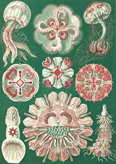
Background imageProtista Collection: Illustration showing a variety of jellyfish. Discomedusae. - Schweibenquallen, 1 print

Illustration showing a variety of jellyfish. Discomedusae. - Schweibenquallen, 1 print : lithograph, color; sheet 36 x 26 cm. 1904

Background imageProtista Collection: Illustration shows starfish and details of starfish anatomy. Ophiodea. - Schlangensterne

Illustration shows starfish and details of starfish anatomy. Ophiodea. - Schlangensterne, 1 print : lithograph; sheet 36 x 26 cm. 1904

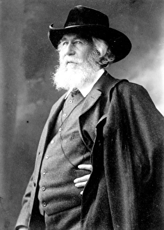
Background imageProtista Collection: Prof. Haeckel, 1 negative : glass; 5 x 7 in

Prof. Haeckel, 1 negative : glass; 5 x 7 in. or smaller. Ernst Haeckel 1834 a 1919 German biologist, naturalist, philosopher, physician, professor, and artist who discovered

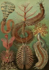
Background imageProtista Collection: Illustration showing a variety of chaetopoda or spined marine worms

Illustration showing a variety of chaetopoda or spined marine worms. Chaetopoda. - Borstenwurmer, 1 print : lithograph, color; sheet 36 x 26 cm. 1904

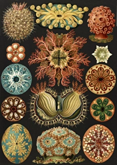
Background imageProtista Collection: Illustration showing a variety of sea squirts. Ascidiae. - Seescheiden, 1 print

Illustration showing a variety of sea squirts. Ascidiae. - Seescheiden, 1 print : lithograph, color; sheet 36 x 26 cm. 1904

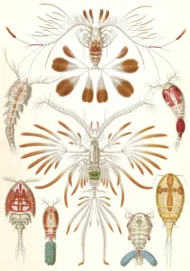
Background imageProtista Collection: Illustration showing a variety of copopod crustaceans. Copepoda. - Ruderkrebse, 1 print

Illustration showing a variety of copopod crustaceans. Copepoda. - Ruderkrebse, 1 print : lithograph, color; sheet 36 x 26 cm. 1904

Background imageProtista Collection: Illustration showing six views of jellyfish. Discomedusae. - Scheibenquallen, 1 print

Illustration showing six views of jellyfish. Discomedusae. - Scheibenquallen, 1 print : lithograph, color; sheet 36 x 26 cm. 1904

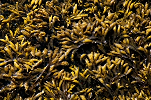
Background imageProtista Collection: Rockweed, Fucus gardneri, Vancouver Island, British Columbia

Rockweed, Fucus gardneri, Vancouver Island, British Columbia

Background imageProtista Collection: Western sandpiper, Calidris mauri, Stanley Park, British Columbia

Western sandpiper, Calidris mauri, Stanley Park, British Columbia

Background imageProtista Collection: Green seaweed

Green seaweed

Background imageProtista Collection: Three dried oyster mushrooms

Three dried oyster mushrooms

Background imageProtista Collection: Large field mushroom and a small button mushroom, view from below

Large field mushroom and a small button mushroom, view from below

Background imageProtista Collection: Scattered button and chestnut mushrooms, close up

Scattered button and chestnut mushrooms, close up

Background imageProtista Collection: Four inverted mushroom caps, close up

Four inverted mushroom caps, close up

Background imageProtista Collection: Suillus lutens, Slippery Jack mushroom emerging from twigs, low angle view

Suillus lutens, Slippery Jack mushroom emerging from twigs, low angle view

Background imageProtista Collection: Spirogyra algae, light micrograph C016 / 9592

Spirogyra algae, light micrograph C016 / 9592
Spirogyra algae. Polarised light micrograph of Spirogyra sp. algae. This filamentous green algae is named for the spiral arrangement of its chloroplasts (green)

Background imageProtista Collection: Spirogyra algae, light micrograph C016 / 9594

Spirogyra algae, light micrograph C016 / 9594
Spirogyra algae. Rheinberg illuminated light micrograph of Spirogyra sp. algae. This filamentous green algae is named for the spiral arrangement of its chloroplasts (green)

Background imageProtista Collection: Desmids and spirogyra, light micrograph C016 / 9595

Desmids and spirogyra, light micrograph C016 / 9595
Desmids and spirogyra. Polarised light micrograph of two Micrasterias rotata desmids (round) and a single filament of Spirogyra sp. green alga (lower right)

Background imageProtista Collection: Spirogyra algae, light micrograph C016 / 9593

Spirogyra algae, light micrograph C016 / 9593
Spirogyra algae. Polarised light micrograph of Spirogyra sp. algae. This filamentous green algae is named for the spiral arrangement of its chloroplasts (green)

Background imageProtista Collection: Fossil foraminifera necklace C016 / 5995

Fossil foraminifera necklace C016 / 5995
Fossil foraminifera (Alveolina sp.) necklace from Sindh, India. The fossils are from Eocene limestone. Necklace from John Whittakers cabinet. Natural History Museum, London, UK

Background imageProtista Collection: Fungus (Lycoperdon coliforme) C016 / 5988

Fungus (Lycoperdon coliforme) C016 / 5988
Fungus (Lycoperdon coliforme). Plate 1 from Drawings of Submerged Algae (1800) by Mary Dawson Turner. Held in the Botany Library at the Natural History Museum, London, UK

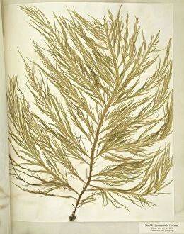
Background imageProtista Collection: Dried seaweed specimens C016 / 5977

Dried seaweed specimens C016 / 5977
Dried seaweed (Chorda filum) specimens. Herbarium sheet from the collection held at the Natural History Museum, London, UK

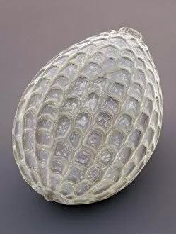
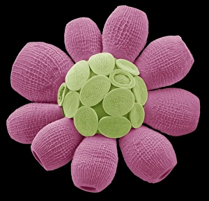
Background imageProtista Collection: Coccolithophore alga, SEM C019 / 0233

Coccolithophore alga, SEM C019 / 0233
Coccolithphore. Coloured scanning electron micrograph (SEM) of part of a coccolithophore unicellular marine alga. It is surrounded by a skeleton (coccosphere) of calcium carbonate plates (coccoliths)

Background imageProtista Collection: Coccolithophore alga, SEM C019 / 0234

Coccolithophore alga, SEM C019 / 0234
Coccolithphore. Coloured scanning electron micrograph (SEM) of part of a coccolithophore unicellular marine alga. It is surrounded by a skeleton (coccosphere) of calcium carbonate plates (coccoliths)

Background imageProtista Collection: Coccolithophore alga, SEM C019 / 0224

Coccolithophore alga, SEM C019 / 0224
Coccolithphore. Coloured scanning electron micrograph (SEM) of a coccolithophore unicellular marine alga. It is surrounded by a skeleton (coccosphere) of calcium carbonate plates (coccoliths)

Background imageProtista Collection: Seaweed (Desmarestia ligulata) C016 / 6014

Seaweed (Desmarestia ligulata) C016 / 6014
seaweed (Desmarestia ligulata). Page 55 from Algae Danmonienses: or dried specimens of Marine Plants, principally collected in Devonshire by Mary Wyatt; carefully named according to Dr

Background imageProtista Collection: Carrageen moss (Chondus cripus) C016 / 6062

Carrageen moss (Chondus cripus) C016 / 6062
Carrageen moss (Chondus cripus). Specimen from Algae Danmonienses: or dried specimens of Marine Plants, principally collected in Devonshire by Mary Wyatt; carefully named according to Dr

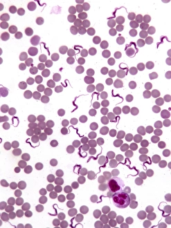
Background imageProtista Collection: Trypanosomes in blood smear, SEM C016 / 5783

Trypanosomes in blood smear, SEM C016 / 5783
Parasitic protozoan. Coloured scanning electron micrograph (SEM) of trypanosome protozoa (Trypanosoma sp.) in a blood smear

Background imageProtista Collection: Foraminiferan, SEM

Foraminiferan, SEM
Foraminiferan. Coloured scanning electron micrograph (SEM) of the foraminiferan Oolina squamosa. Foraminiferans are marine single-celled protists that construct and inhabit shells (tests)

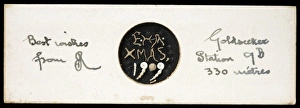
Background imageProtista Collection: Heron- Allen microscope slides of foraminifera

Heron- Allen microscope slides of foraminifera
Christmas greetings spelt out in minute shells which bears the initials of the maker, Edward Heron-Allen, and the year it was made: E H A, 1909

Background imageProtista Collection: Scyphosphaera apsteinii

Scyphosphaera apsteinii. SEM image of an equatorial coccolith

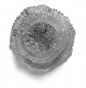
Background imageProtista Collection: Model of coccolith Kamptnerius magnificus

Model of coccolith Kamptnerius magnificus
Copy of the resin model of the coccolith Kamptnerius magnificus Deflandre, 1930 currently on display in the Earth Lab area of the Earth Galleries at the Natural History Museum, London


All Professionally Made to Order for Quick Shipping

"Exploring the Intricate World of Protista: From Pressed Seaweed Specimens to Microscopic Wonders" In this captivating journey through the diverse realm of protista, we encounter a fascinating array of organisms. The pressed seaweed specimens C016 / 6127 reveal intricate patterns and textures that hint at the hidden beauty within. Diatoms, as seen under a scanning electron microscope (SEM), showcase their mesmerizing geometric shapes and delicate structures. Dictyota dichotoma, a brown algae commonly known as mermaid's fan, unveils its elegant fronds swaying in underwater currents. Illustrations depicting hummingbirds from the Trochilidae family remind us of the interconnectedness between these avian wonders and protists. These tiny creatures rely on nectar produced by flowering plants like Fucus bulbosus, a type of kelp found along coastal regions. Another diatom specimen captured under SEM reveals its intricate details, highlighting nature's artistry at microscopic scales. Fucus radiatus, another species of kelp with branching thalli resembling miniature forests beneath the waves, showcases its resilience in harsh marine environments. Moving beyond seaweeds and birds, illustrations introduce us to liverworts from the Hepaticae family - primitive land-dwelling plants that share an ancient lineage with protists. Their unique reproductive strategies have allowed them to thrive for millions of years. However, not all protists are benign or beneficial; some can cause harm. Plasmodium sp. , a malarial parasite transmitted by mosquitoes, serves as a stark reminder of how these microorganisms can impact human health. Radiolarians belonging to Acanthophracta group astonish with their intricate skeletal structures made up of silica shells. Coelosphaeridium presents yet another marvel – calcareous alga adorned with ornate patterns reminiscent of stained glass windows. Throughout this exploration into the world of protista, diatoms emerge as prominent protagonists.