
Protein Coat Collection
"The Intricate World of Protein Coats: Unveiling the Hidden Guardians" Protein coats, also known as viral capsids


All Professionally Made to Order for Quick Shipping
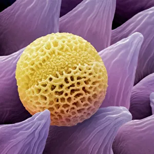
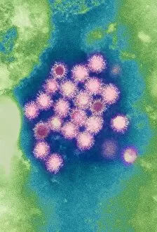
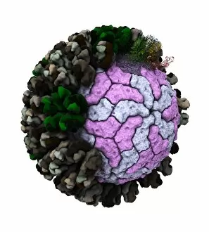
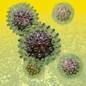
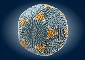
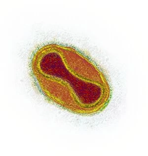
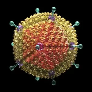
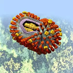
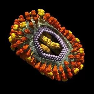
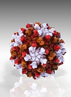
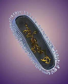
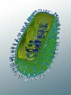
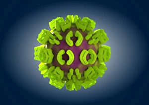
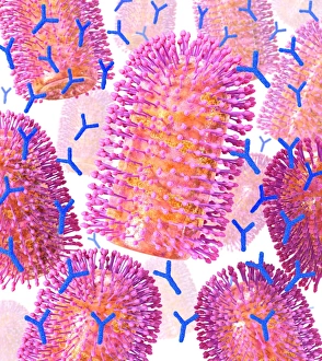
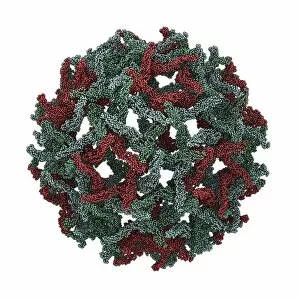
"The Intricate World of Protein Coats: Unveiling the Hidden Guardians" Protein coats, also known as viral capsids, are nature's ingenious armor that safeguards a variety of microscopic entities. These captivating structures play a pivotal role in protecting and delivering genetic material within viruses. Let us embark on an enlightening journey into their mesmerizing realm. Intricately detailed under the scanning electron microscope (SEM), lavender pollen grains reveal their protein coat's remarkable architecture. Like tiny fortresses, these coats shield the precious DNA or RNA cargo from external threats while ensuring successful pollination. Moving onto more menacing inhabitants, hepatitis B viruses showcase their distinctive protein coats through stunning artwork representations. The intricate patterns and shapes depicted highlight the virus's ability to evade our immune system by camouflaging itself with this protective layer. Rotavirus particles captivate us with artistic renderings showcasing their spherical shape enveloped by a robust protein coat. This armor shields its genome during transmission, enabling it to cause gastrointestinal distress in unfortunate hosts. Delving further into hepatitis B virus particles' artwork reveals diverse variations in their protein coatings – C016 / 9097, C016 / 9129, and C016 / 9126. Each unique pattern represents different strains of this notorious virus responsible for liver infections worldwide. Parvovirus particle artwork (C013 / 4640) unravels yet another facet of these fascinating structures. Their symmetrical arrangement exemplifies how proteins assemble meticulously around viral genomes to ensure efficient replication and infection processes. The poliovirus particle F006 / 9306 showcases its iconic appearance encapsulated within its characteristic protein coat. This visual representation reminds us of humanity's triumph over one of history's most devastating diseases through vaccination campaigns targeting this very structure. Not limited to pathogenic viruses alone, human adenovirus 36 (artwork C016 / 8966) demonstrates how even benign viruses employ elaborate protein coats for their survival.