
Posterior View Collection
"Exploring the Posterior View: From Stretching to Anatomy" A woman stretching her body, showcasing the posterior view and promoting flexibility


All Professionally Made to Order for Quick Shipping
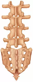
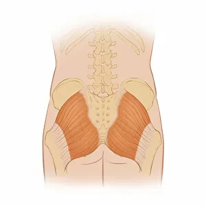
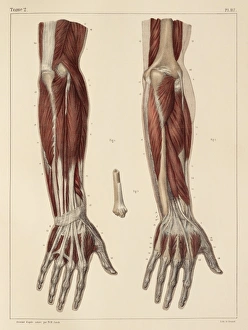
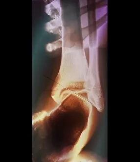
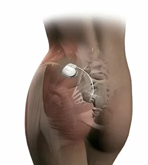
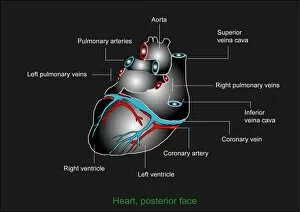
"Exploring the Posterior View: From Stretching to Anatomy" A woman stretching her body, showcasing the posterior view and promoting flexibility. Delving into the intricate details of a normal heart and its arteries from a posterior perspective. An enchanting engraving from 1914 reveals an anatomical view of the human torso, emphasizing its posterior aspects. Travel back in time with a captivating screen print from 1935, displaying an anatomical print of the human skeleton and muscles seen from behind. Witnessing medical precision, observe a posterior view of the patellar surface featuring healthy cartilage functioning normally. Unveiling medical intervention, witness a shaver delicately cleaning up injured cartilage on the posterior view of the patellar surface. Gaining insight into spinal health, explore a normal posterior view of both lumbar spine and sacrum structures for better understanding. Discovering strength in motion, admire the gluteus maximus muscle alongside hip bones in their natural state through a normal posterior perspective. Appreciating historical artwork's contribution to anatomy education; marvel at detailed illustrations showcasing forearm muscles dating back to 1831. Examining injury management techniques, delve into another glimpse of injured cartilage on the patellar surface through its distinctive rear aspect. Admiring artistic mastery while learning about superficial back muscles through an exquisite artwork created in 1831 Unraveling breathing mechanics with elegance.