Fraud Blocker Skip to main content

Portal Vein Collection

Exploring the intricacies of human anatomy, let's delve into the role of the portal vein

Background imagePortal Vein Collection: Liver cirrhosis

Liver cirrhosis. Artwork taken from Sir Robert Carswells textbook Pathological Anatomy, published 1838. Cirrhosis is a disease in which the liver tissue is replaced with scar tissue

Background imagePortal Vein Collection: Human male anatomy

Human male anatomy
Antique illustration of a Human male anatomy

Background imagePortal Vein Collection: Portal vein surgery, X-ray C016 / 6546

Portal vein surgery, X-ray C016 / 6546
Vein surgery. Coloured angiogram (X-ray) of surgery to lower the blood pressure in the portal vein of a 60 year old patient

Background imagePortal Vein Collection: Portal vein surgery, X-ray C016 / 6543

Portal vein surgery, X-ray C016 / 6543
Vein surgery. Angiogram (X-ray) of surgery to lower the blood pressure in the portal vein of a 60 year old patient. A catheter (white line running down from top centre)

Background imagePortal Vein Collection: Portal vein surgery, X-ray C016 / 6545

Portal vein surgery, X-ray C016 / 6545
Vein surgery. Coloured angiogram (X-ray) of surgery to lower the blood pressure in the portal vein of a 60 year old patient

Background imagePortal Vein Collection: Portal vein surgery, X-ray C016 / 6544

Portal vein surgery, X-ray C016 / 6544
Vein surgery. Coloured angiogram (X-ray) of surgery to lower the blood pressure in the portal vein of a 60 year old patient

Background imagePortal Vein Collection: Liver and stomach arteries, 1825 artwork

Liver and stomach arteries, 1825 artwork
Liver and stomach arteries. Dissection of the abdomen, showing the arteries (red) of the liver (brown, upper left), stomach (pink, upper centre), and the intestines (grey with yellow fat)

Background imagePortal Vein Collection: Veins of the stomach, 1825 artwork

Veins of the stomach, 1825 artwork
Veins of the stomach. Abdominal dissection showing the veins (blue) of the stomach (centre) and the greater omentum (lower centre, yellow)

Background imagePortal Vein Collection: False-colour SEM of vessels in liver lobule

False-colour SEM of vessels in liver lobule
Liver tissue. False-colour scanning electron micrograph (SEM) of blood vessels around a lobule, the functional unit of the liver

Background imagePortal Vein Collection: Stem cell therapy, artwork

Stem cell therapy, artwork
Stem cell therapy. Artwork showing stem cells (in green cylinder) being added to a patients bloodstream in the region of the liver (brown)


All Professionally Made to Order for Quick Shipping

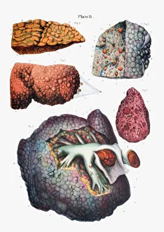
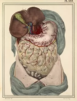
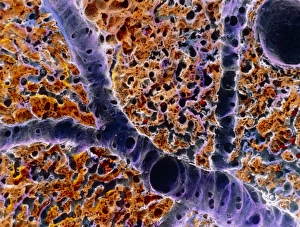
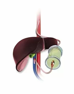
Exploring the intricacies of human anatomy, let's delve into the role of the portal vein. This vital blood vessel carries blood from the digestive system, including the stomach and pancreas, to the liver. In cases of liver cirrhosis, the portal vein may become obstructed, leading to potential complications. Portal vein surgery, as depicted in X-rays C016 / 6543, 6544, 6545, and 6546, can help alleviate these issues. The ancient 1825 artwork "Liver and Stomach Arteries" offers a historical perspective, while modern technology, such as false-color SEM images of vessels in a liver lobule, reveals the intricate details. The future treatment may lie in stem cell therapy, as depicted in artistic renderings, offering new possibilities for healing and restoration.