
Polarised Light Microscope Collection
The polarised light microscope is a powerful tool that allows scientists to delve into the intricate world of crystals and microorganisms


All Professionally Made to Order for Quick Shipping
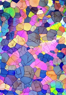
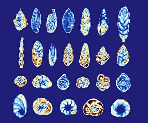
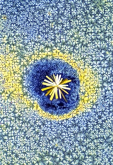
The polarised light microscope is a powerful tool that allows scientists to delve into the intricate world of crystals and microorganisms. With its ability to reveal hidden details, it uncovers mesmerizing patterns and structures that are otherwise invisible to the naked eye. In one captivating image, caffeine crystals come alive under the lens, showcasing their delicate arrangement in a mesmerizing dance of symmetry. The light micrograph captures their beauty, highlighting the intricate network they form. Moving on to another fascinating subject, cortisol crystals take center stage in yet another light micrograph. Their unique formation resembles an otherworldly landscape, with jagged edges and interlocking shapes creating an ethereal visual experience. Enkephalin crystals also make an appearance in this captivating collection of images. Through the lens of the polarised light microscope, these tiny formations reveal themselves as intricately woven threads of brilliance. Cortisol crystals return once more but with a twist - captured in a different angle or lighting condition (C015 / 6786). This variation showcases how even slight changes can transform our perception of these microscopic wonders. Foraminiferans add diversity to this stunning series of photographs. These marine organisms appear like delicate shells suspended within time, each telling its own story through its unique structure and design. GABA crystals bring forth a new dimension as they shimmer under polarised light. Their radiant display captivates viewers with their vibrant colors and geometric precision. Vitamin B1 and Vitamin B7 crystals grace us with their presence next – two essential nutrients transformed into dazzling works of art when viewed through this remarkable microscope. The symmetrical arrangements evoke feelings of harmony and balance within nature's building blocks. Lastly, adenosine crystals join forces with GABA for one final spectacle before concluding this extraordinary journey through the polarised light microscope's lens. Together they create a breathtaking display that merges elegance with complexity – reminding us once again just how awe-inspiring our microscopic world can be.