Fraud Blocker Skip to main content

Polarised Light Micrograph Collection (page 3)

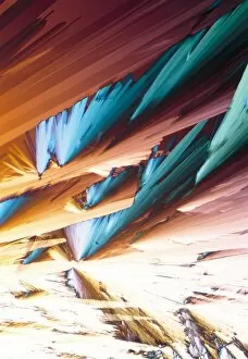
Capturing the mesmerizing beauty of microscopic crystals through polarised light micrography is truly a sight to behold

Background imagePolarised Light Micrograph Collection: Vitamin B7 crystals, light micrograph

Vitamin B7 crystals, light micrograph
Vitamin B7 crystals, polarised light micrograph. Vitamin B7, also called biotin or vitamin H, is an essential nutrient used by the body in cell growth

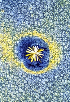
Background imagePolarised Light Micrograph Collection: Adenosine crystals, light micrograph

Adenosine crystals, light micrograph
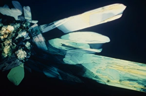
Adenosine crystals, polarised light micrograph. Adenosine is a nucleoside molecule composed of the base adenine bonded to the sugar ribose

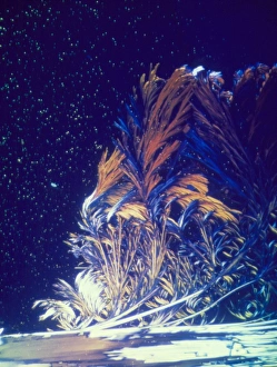
Background imagePolarised Light Micrograph Collection: Enkephalin crystals, light micrograph

Enkephalin crystals, light micrograph
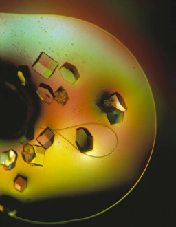
Enkephalin crystals, polarised light micrograph. Enkephalin is an endorphin found in the human brain. There are two variants: Met-enkephalin (seen here), which contains the amino acid methionine

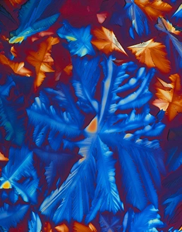
Background imagePolarised Light Micrograph Collection: Vitamin B1 crystals, light micrograph

Vitamin B1 crystals, light micrograph
Vitamin B1 crystals, polarised light micrograph. Vitamin B1, also called thiamine, is an essential nutrient that humans are unable to produce and so need to obtain from their diet

Background imagePolarised Light Micrograph Collection: Cholecystokinin crystals

Cholecystokinin crystals, polarised light micrograph. Cholecystokinin (CCK) is a hormone responsible for promoting the digestion of fats and proteins

Background imagePolarised Light Micrograph Collection: Glutamic acid crystals, light micrograph

Glutamic acid crystals, light micrograph
Glutamic acid crystals, polarised light micrograph. Glutamic acid is an non-essential amino acid, in which it can be synthesised by the body and does not need to be obtained from food

Background imagePolarised Light Micrograph Collection: Cortisol crystals, light micrograph

Cortisol crystals, light micrograph
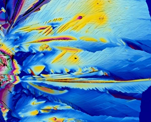
Cortisol crystals, polarised light micrograph. Cortisol is a steroid hormone produced by the adrenal glands, which sit on top of the kidneys

Background imagePolarised Light Micrograph Collection: GABA crystals, light micrograph

GABA crystals, light micrograph
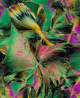
GABA crystals. Polarised light micrograph of gamma-aminobutyric acid (GABA). GABA is an inhibitory neurotransmitter (nerve signalling chemical) that regulates the excitability of all nerve cells

Background imagePolarised Light Micrograph Collection: Acetylcholine crystals, light micrograph

Acetylcholine crystals, light micrograph
Acetylcholine crystals, polarised light micrograph. Acetylcholine (ACh) is a neurotransmitter (nerve signalling chemical) that plays an important role in relaying impulses at myoneural (muscle-nerve)

Background imagePolarised Light Micrograph Collection: Retinoic acid crystal, light micrograph

Retinoic acid crystal, light micrograph
Retinoic acid crystal, polarised light micrograph. Retinoic acid is one of three forms of vitamin A; the other two are retinol and retinal

Background imagePolarised Light Micrograph Collection: Cholecystokinin crystal

Cholecystokinin crystal, polarised light micrograph. Cholecystokinin (CCK) is a hormone responsible for promoting the digestion of fats and proteins

Background imagePolarised Light Micrograph Collection: NMDA crystals, light micrograph

NMDA crystals, light micrograph
NMDA crystals, polarised light micrograph. NMDA (N-methyl-D-aspartic acid) is an amino acid derivative and a neurotransmitter (nerve signalling chemical)

Background imagePolarised Light Micrograph Collection: Immunoglobulin crystals, light micrograph

Immunoglobulin crystals, light micrograph
Immunoglobulin crystals, polarised light micrograph. Immunoglobulins, or antibodies, are a type of globulin protein that are produced by B cells (a type of white blood cell)

Background imagePolarised Light Micrograph Collection: Butchers broom stem, light micrograph

Butchers broom stem, light micrograph
Butchers broom stem. Polarised light micrograph of a section through a butchers broom stem (Ruscus aculeatis) showing many vascular bundles (dark clusters) within the plant cortex (orange)

Background imagePolarised Light Micrograph Collection: Umbrella grass stem, light micrograph

Umbrella grass stem, light micrograph
Umbrella grass stem. Polarised light micrograph of a section through a umbrella grass stem (Cyperus alternifolius). Vascular bundles (brown) can be seen containing xylem (larger openings)

Background imagePolarised Light Micrograph Collection: Purple moor grass stem, light micrograph

Purple moor grass stem, light micrograph
Purple moor grass stem. Polarised light micrograph of a section through a purple moor grass stem (Molinia caerula). Vascular bundles (round orange structures)

Background imagePolarised Light Micrograph Collection: Bamboo stem, light micrograph

Bamboo stem, light micrograph
Bamboo stem. Polarised light micrograph of a section through a bamboo stem (Bambusa vulgaris). Vascular bundles can be seen (yellow/green) containing xylem (larger openings)

Background imagePolarised Light Micrograph Collection: Maize stem, light micrograph

Maize stem, light micrograph
Maize stem. Polarised light micrograph of a section through a maize plant stem (Zea mays). A vascular bundles (brown) can be seen containing xylem (larger openings)

Background imagePolarised Light Micrograph Collection: Spiny rush stem, light micrograph

Spiny rush stem, light micrograph
Spiny rush stem. Polarised light micrograph of a section through a spiny rush stem (Juncus actus). Vascular bundles can be seen (purple/red) containing xylem (larger openings)

Background imagePolarised Light Micrograph Collection: Sugar cane stem, light micrograph

Sugar cane stem, light micrograph
Sugar cane stem. Polarised light micrograph of a section through a sugar cane stem (Saccharum officinareum). Vascular bundles (round blue/purple structures)

Background imagePolarised Light Micrograph Collection: Sarsaparilla plant stem, light micrograph

Sarsaparilla plant stem, light micrograph
Maize root. Polarised light micrograph of a section through the root of a sarsaparilla plant (Smilax regelii) showing a vascular cylinder

Background imagePolarised Light Micrograph Collection: Inulin crystals, light micrograph

Inulin crystals, light micrograph
Inulin crystals. Polarised light micrograph of inulin crystals (yellow) stored inside a Dahlia sp. root tuber. Inulin is a naturally-occurring plant sugar found in the roots or rhizomes of some

Background imagePolarised Light Micrograph Collection: Black burdock stem, light micrograph

Black burdock stem, light micrograph
Black burdock stem. Polarised light micrograph of a transverse section through the stem of a black burdock (Arctium lappa) plant

Background imagePolarised Light Micrograph Collection: Stinging nettle leaf, light micrograph

Stinging nettle leaf, light micrograph
Stinging nettle leaf. Polarised light micrograph of the underside of a stinging nettle (Urtica dioica) leaf. Two types of hairs are seen

Background imagePolarised Light Micrograph Collection: Sunflower root, light micrograph

Sunflower root, light micrograph
Sunflower root. Polarised light micrograph of a transverse section through the root of a sunflower (Helianthus annuus) plant

Background imagePolarised Light Micrograph Collection: Orchid starch grains, light micrograph

Orchid starch grains, light micrograph
Orchid starch grains. Polarised light micrograph of a transverse section through an orchid tuber (Phaius grandifolius), showing the starch grains

Background imagePolarised Light Micrograph Collection: Dodder parasitic plant

Dodder parasitic plant. Polarised light micrograph of a section through a host plant parasitised by a dodder (Cuscuta sp.)

Background imagePolarised Light Micrograph Collection: Rhubarb leaf, light micrograph

Rhubarb leaf, light micrograph
Rhubarb leaf. Polarised light micrograph of a longitudinal section through a rhubarb (Rheum sp.) leaf. The dark blue structure is a vascular bundle

Background imagePolarised Light Micrograph Collection: Elm stem, light micrograph

Elm stem, light micrograph
Elm stem (Ulmus procera) section, polarised light micrograph. The black horizontal lines are multiseriate rays, which are composed of several rows of cells

Background imagePolarised Light Micrograph Collection: Black pepper stem, light micrograph

Black pepper stem, light micrograph
Black pepper stem. Polarised light micrograph of a transverse section through the stem of a black pepper (Pipa nigrum) plant

Background imagePolarised Light Micrograph Collection: Paracetamol crystals, light micrograph

Paracetamol crystals, light micrograph
Paracetamol. Polarised light micrograph of paracetamol (acetaminophen) crystals. Paracetamol is an analgesic (pain killer) and antipyretic (reduces fever)

Background imagePolarised Light Micrograph Collection: Light micrograph of arginine crystals

Light micrograph of arginine crystals
Polarised light micrograph of crystals of the amino acid arginine (ARG). There are twenty standard amino acids, which form the basic building blocks of all living matter

Background imagePolarised Light Micrograph Collection: Co-enzyme NAD (nicotinamide) crystals

Co-enzyme NAD (nicotinamide) crystals
Polarised light micrograph of crystals of the co- enzyme nicotinamide adenine dinucleotide (NAD). This is an extremely important and widespread co- enzyme found in nature

Background imagePolarised Light Micrograph Collection: Vitamin C crystals, PLM

Vitamin C crystals, PLM
Vitamin C crystals, polarised light micrograph. Vitamin C (ascorbic acid) is a chemical essential for good health. It is required for the production of collagen

Background imagePolarised Light Micrograph Collection: Polarised LM of vitamin D crystals

Polarised LM of vitamin D crystals
Vitamin D. Polarised light micrograph of crystals of vitamin D. Vitamin D is fat-soluble, and is found in foods such as animal fats, milk, butter and eggs

Background imagePolarised Light Micrograph Collection: Light micrograph of methionine crystals

Light micrograph of methionine crystals
Polarised light micrograph of crystalline Methionine. Magnification: x40 at 35mm size

Background imagePolarised Light Micrograph Collection: Lysozome protein crystals

Lysozome protein crystals
Lysozyme enzyme crystals. Polarised light micro- graph of crystals of the enzyme lysozyme from the egg white (albumen) of a domestic chicken

Background imagePolarised Light Micrograph Collection: Folic acid crystals

Folic acid crystals

Background imagePolarised Light Micrograph Collection: Light micrograph of thiamine crystals

Light micrograph of thiamine crystals

Background imagePolarised Light Micrograph Collection: Vitamin B3 crystals

Vitamin B3 crystals
Vitamin B3. Polarised light micrograph of vitamin B3 crystals. Vitamin B3 exists in two main forms: niacin and niacinamide

Background imagePolarised Light Micrograph Collection: Copper sulphate crystals, PLM

Copper sulphate crystals, PLM
Copper sulphate crystals, polarised light micrograph. This is hydrated copper (II) sulphate, which has the chemical formula CuSO4.5H2O. On a macroscopic scale, the crystals are a bright, intense blue

Background imagePolarised Light Micrograph Collection: Polarised LM of niacinamide crystals

Polarised LM of niacinamide crystals
Nicotinamide. Polarised light micrograph of crystals of nicotinamide, or niacinamide. This is the amide of nicotinic acid. The acid and amide are both B vitamins, and are equally active

Background imagePolarised Light Micrograph Collection: PLM of crystals of pantothenic acid

PLM of crystals of pantothenic acid
Pantothenic acid. Polarised light micrograph of crystals of pantothenic acid, a vitamin of the B- complex group of vitamins. Magnification: x63 at 35mm size

Background imagePolarised Light Micrograph Collection: Ascorbic and citric acid crystals, PLM

Ascorbic and citric acid crystals, PLM
Citric and ascorbic acid crystals, polarised light micrograph. Ascorbic acid, also known as vitamin C, is a nutrient essential for health

Background imagePolarised Light Micrograph Collection: Cholesterol crystals

Cholesterol crystals
Cholesterol. Light micrograph obtained with polarised light of crystals of cholesterol. It is a lipid (fatty substance) found in vertebrates and in foods from animal sources

Background imagePolarised Light Micrograph Collection: Nylon 6 crystals

Nylon 6 crystals
Nylon crystals. Polarised light micrograph of nylon 6 spherulites (crystals). The spherulites appear as yellow and blue circles in a cross pattern

Background imagePolarised Light Micrograph Collection: Plant stem trichomes, light micrograph

Plant stem trichomes, light micrograph
Plant stem trichomes. Polarised light micrograph of simple multicellular hairs (trichomes) on the surface (epidermis) of a stem of hedge woundwort (Stachys sylvatica)

Background imagePolarised Light Micrograph Collection: Tree xylem vessels, light micrograph

Tree xylem vessels, light micrograph
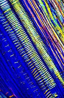
Tree xylem vessels. Polarised light micrograph of a vertical section through the primary wood vessels (xylem vessels) of a tree


All Professionally Made to Order for Quick Shipping

Capturing the mesmerizing beauty of microscopic crystals through polarised light micrography is truly a sight to behold. In this captivating world, caffeine crystals take center stage, showcasing their intricate patterns and delicate structure under the lens. The vibrant hues and symmetrical arrangements of these caffeine crystals create a visual spectacle that leaves us in awe. Moving on from caffeine, oxytocin hormone crystals make their appearance in PLM C016 / 7196, revealing an enchanting display of shimmering facets. These tiny wonders hold within them the power to evoke feelings of love and bonding. Similarly, oxytocin crystals captured in another light micrograph unveil their breathtaking elegance as they glisten with ethereal radiance. Cortisol crystals also join the lineup, offering a glimpse into their unique composition through yet another stunning light micrograph. Their sharp edges and intricate formations remind us of the complex nature of stress hormones present within our bodies. Returning to caffeine once again, its crystalline form takes on new dimensions when observed under polarised light microscopy. The interplay between colors and shapes creates an otherworldly landscape that transports us into a realm where science meets art. Testosterone makes its grand entrance through PLM crystal imagery, displaying its own distinct patterned arrangement that reflects strength and vitality. This snapshot allows us to appreciate the intricacies hidden within this essential hormone for both men and women alike. Insulin crystals come alive in yet another mesmerizing light micrograph (C017 / 8249), showcasing their delicate symmetry while reminding us of their vital role in regulating blood sugar levels within our bodies. Oxytocin hormone returns once more (LM C016 / 7195), captivating our imagination with its alluring crystal formation that hints at deep emotional connections we share as humans - bonds formed by trust, empathy, and compassion. Amidst these crystalline marvels lies a water fern rhizome captured under bright illumination.