
Plant Pathology Collection
"Exploring the Intricate World of Plant Pathology: Unveiling the Secrets of Disease" In the realm of plant pathology


All Professionally Made to Order for Quick Shipping
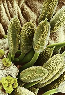
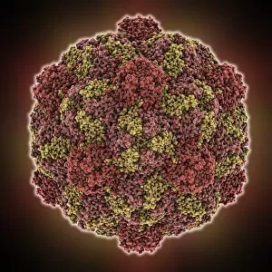
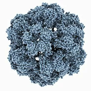
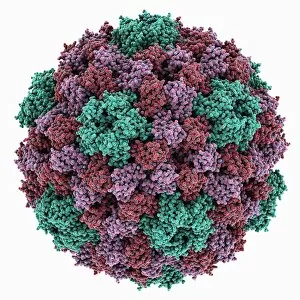
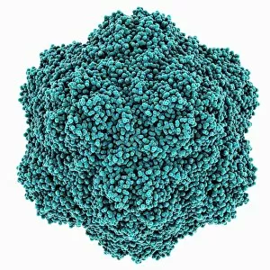
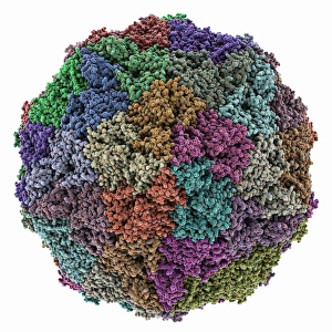
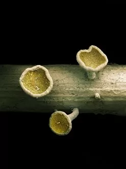
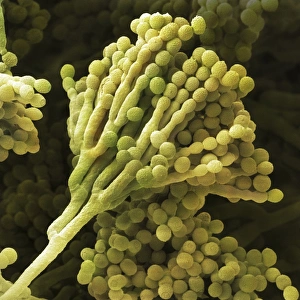
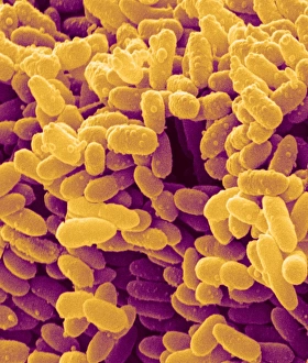
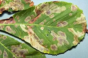
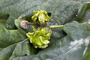
"Exploring the Intricate World of Plant Pathology: Unveiling the Secrets of Disease" In the realm of plant pathology, nature's battles unfold as microscopic warriors engage in a constant struggle for survival. From bleeding cankers to rust fungi on rose leaves, these captivating images captured by scanning electron microscopy (SEM) offer a glimpse into this hidden world. Behold the intricate beauty of turnip yellow mosaic virus capsid and brome mosaic virus capsid, resembling delicate works of art that belie their destructive potential. The cowpea chlorotic mottle virus capsid and tobacco necrosis virus capsid showcase stunning geometric patterns, reminding us that even in chaos, there is order. Venturing deeper into this unseen realm, we encounter the grapevine fanleaf virus capsid – an enigmatic structure embodying both fragility and resilience. Meanwhile, root-knot nematode larvae lurk beneath the soil surface, their presence causing havoc to plant roots. Rust fungus on rose leaves becomes an artistic masterpiece under SEM's lens - its spores intricately arranged like a painter's brushstrokes across a canvas. These captivating images serve as reminders that beauty can be found even amidst destruction. Amidst these visual wonders lies ongoing research on citrus greening disease – scientists tirelessly striving to unravel its mysteries and find solutions to protect our beloved citrus trees from devastation. As we delve further into this mesmerizing world through SEM imagery, it becomes evident that plant pathology is not merely about studying diseases; it is about understanding complex interactions between plants and pathogens, and is about safeguarding our crops' health while preserving biodiversity for future generations. So let us marvel at these snapshots frozen in time – each image telling a story of resilience or vulnerability within the intricate web woven by nature herself. In embracing plant pathology's challenges head-on, we unlock crucial knowledge vital for sustaining our planet's green tapestry.