Fraud Blocker Skip to main content

Plant Cell Collection

"Exploring the Intricate World of Plant Cells

Background imagePlant Cell Collection: Plant cell

Plant cell
False-colour transmission electron micrograph (TEM) of a cell in the root tip of a maize plant, Zea mays. Plant cells are distinct from animal cells in having an additional external envelope

Background imagePlant Cell Collection: Cell types, artwork

Cell types, artwork
Cell types. Cutaway artwork with a scale bar (upper right, in micrometres) showing the relative sizes of eukaryotic cells (those containing a nucleus) and prokaryotic cells (those lacking a nucleus)

Background imagePlant Cell Collection: Chloroplast structure, artwork

Chloroplast structure, artwork
Chloroplast structure. Artwork showing the internal structure of chloroplasts, the organelles in plant cells responsible for photosynthesis

Background imagePlant Cell Collection: Illustration of cross-section through leaf, showing upper epidermis, palisade mesophyll, xylem vesse

Illustration of cross-section through leaf, showing upper epidermis, palisade mesophyll, xylem vesse

Background imagePlant Cell Collection: Golgi apparatus, TEM C017 / 8234

Golgi apparatus, TEM C017 / 8234
Golgi apparatus. Transmission electron micrograph (TEM) of Golgi apparatus from a Chlamydomonas sp. alga. Golgi are membrane-bound organelles that modify and package proteins

Background imagePlant Cell Collection: Tobacco necrosis virus research, artwork

Tobacco necrosis virus research, artwork
Tobacco necrosis virus research, computer artwork

Background imagePlant Cell Collection: Illustration of the structure of a plant cell, including nucleus, nucleolus, ribosome, endoplasmatic

Illustration of the structure of a plant cell, including nucleus, nucleolus, ribosome, endoplasmatic

Background imagePlant Cell Collection: Illustration of different types of cells, nerve cells, red blood cell, muscle cell

Illustration of different types of cells, nerve cells, red blood cell, muscle cell, and guard cells of a leaf

Background imagePlant Cell Collection: Illustration of the life cycle of a fern, inset showing spermatozoa entering female organ

Illustration of the life cycle of a fern, inset showing spermatozoa entering female organ (archegonia)

Background imagePlant Cell Collection: Illustration of leaf anatomy

Illustration of leaf anatomy

Background imagePlant Cell Collection: Micrograph of onion cells, close-up

Micrograph of onion cells, close-up

Background imagePlant Cell Collection: Micrograph of cross-section through a monocotyledonous root (maize)

Micrograph of cross-section through a monocotyledonous root (maize)

Background imagePlant Cell Collection: Chloroplast, artwork

Chloroplast, artwork
Chloroplast, computer artwork

Background imagePlant Cell Collection: Plant cell, artwork F006 / 9876

Plant cell, artwork F006 / 9876
Plant cell, computer artwork

Background imagePlant Cell Collection: Mitochondrion structure, artwork

Mitochondrion structure, artwork
Mitochondrion structure. Artwork showing the internal structure of a mitochondrion. This structure, found in eukaryotic cells, is the site of energy production

Background imagePlant Cell Collection: Mitosis in root tip of Broad Bean, LM

Mitosis in root tip of Broad Bean, LM
Light micrograph (longitudinal section) of the cells in the root tip of a broad bean plant Vicia faba. The dark spot in the centre of each cell is the nucleus

Background imagePlant Cell Collection: Plant cells, artwork

Plant cells, artwork
Plant cells, computer artwork

Background imagePlant Cell Collection: Marsh marigold carpel, light micrograph

Marsh marigold carpel, light micrograph
Marsh marigold carpel. Light micrograph of a transverse section through the carpel of a marsh marigold (Caltha palustris) flower. The ovules (dark green) contain meiotic embryonic cells (brown)

Background imagePlant Cell Collection: Plant cell, artwork

Plant cell, artwork
Plant cell, computer artwork

Background imagePlant Cell Collection: Plant evolution

Plant evolution. Conceptual computer artwork of plant evolution. Two molecules of DNA (deoxyribonucleic acid) are emerging from plant cells (bottom) and joining together (upper centre)

Background imagePlant Cell Collection: Chloroplasts, light micrograph

Chloroplasts, light micrograph. Chloroplasts are one of the features that distinguish a plant cell from an animal cell. They contain chlorophyll

Background imagePlant Cell Collection: Plant cells, light micrograph

Plant cells, light micrograph. These cells are from a spiderwort (Tradescantia) plant. The rigid shape of a plants cell wall is one of the features that distinguishes it from animal cells

Background imagePlant Cell Collection: Cytokinesis, artwork

Cytokinesis, artwork
Cytokinesis. Artwork showing the stage of cell division that involves the splitting of the cell cytoplasm between two daughter cells

Background imagePlant Cell Collection: Cytokinesis, diagram

Cytokinesis, diagram
Cytokinesis. Diagram showing the stage of cell division that involves the splitting of the cell cytoplasm between two daughter cells

Background imagePlant Cell Collection: Plant cell, SEM

Plant cell, SEM
Plant cell. Coloured scanning electron micrograph (SEM) of a section through a plant cell, revealing its internal structure. The cell is encased in a cellulose, hemicellulose and pectin cell wall

Background imagePlant Cell Collection: Moss leaf cells, light micrograph

Moss leaf cells, light micrograph
Moss leaf cells. Light micrograph of cells in a leaf of shining Hookeria moss (Hookeria luscens). Two whole cells (horizontal) are seen at centre, surrounded by parts of other cells

Background imagePlant Cell Collection: Rosemary leaf, light micrograph

Rosemary leaf, light micrograph
Rosemary leaf. Light micrograph of a transverse section through a rosemary (Romarinus officinalis) leaf. The leaf of this plant has a number of adaptations that help it to minimise water loss through

Background imagePlant Cell Collection: Pincushion leaf, light micrograph

Pincushion leaf, light micrograph
Pin cushion leaf. Polarised light micrograph of a transverse section through a pinchusion (Hakea laurina) leaf. The leaf of this plant has a number of adaptations that help it to minimise water loss


All Professionally Made to Order for Quick Shipping

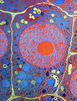
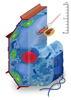
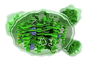
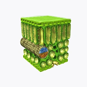
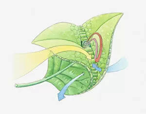
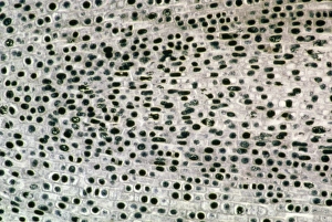
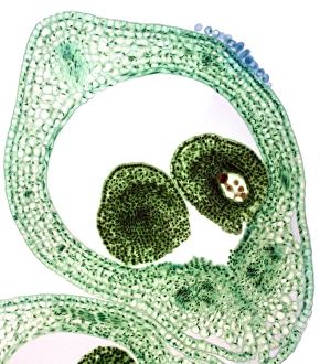
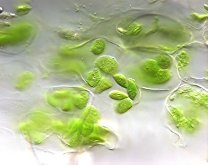
"Exploring the Intricate World of Plant Cells: A Visual Journey" Step into the fascinating realm of plant cells as we delve into their diverse types and intricate structures through captivating artwork. Marvel at the mesmerizing chloroplast structure, depicted in stunning detail, where photosynthesis takes place, harnessing sunlight to produce energy for plants. Observe a magnificently illustrated plant cell, with its nucleus, nucleolus, ribosomes, and endoplasmic reticulum intricately portrayed. Discover the vital components that enable these cells to carry out essential functions within plants. Venture further into our exploration as we unveil different cell types such as nerve cells, red blood cells, muscle cells, and even guard cells found in leaves. Witness how each type plays a unique role in maintaining plant health and functionality. Embark on an enlightening journey through tobacco necrosis virus research artwork that sheds light on scientific breakthroughs aiming to combat this destructive pathogen affecting plants worldwide. Immerse yourself in an illustration showcasing a cross-section through a leaf's upper epidermis alongside palisade mesophyll and xylem vessels. Gain insights into how these layers contribute to leaf function and overall plant vitality. Delve deeper into nature's wonders with an illustration depicting the life cycle of a fern – witness spermatozoa entering female organs (archegonia) during reproduction. Marvel at the complexity of nature's reproductive processes. Explore leaf anatomy like never before with detailed illustrations unraveling its inner workings - from stomata regulating gas exchange to veins transporting water and nutrients throughout the leaf structure. Zoom in on onion cells captured under high-resolution micrographs revealing their intricate patterns up close – marvel at their beauty hidden beneath everyday vegetables. Finally, take a microscopic glimpse into monocotyledonous root structure by examining cross-sections of maize roots under intense magnification – uncovering secrets hidden beneath Earth's surface.