
Pineal Gland Collection
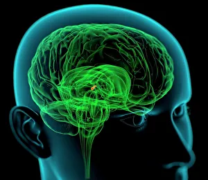
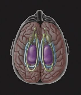
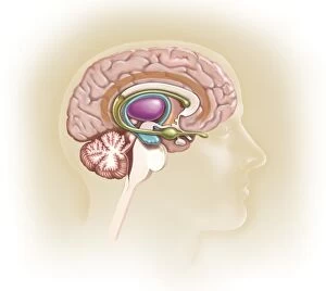
The pineal gland, located in the medulla oblongata of the brain, has long fascinated scientists and philosophers alike


All Professionally Made to Order for Quick Shipping
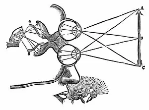
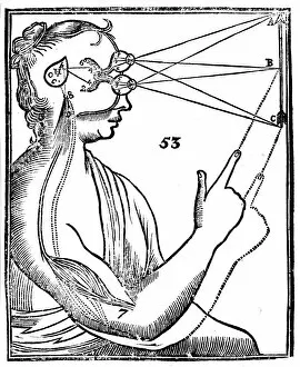
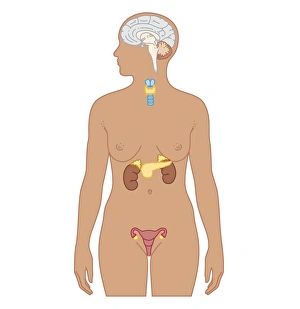
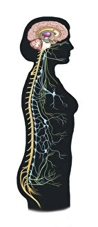
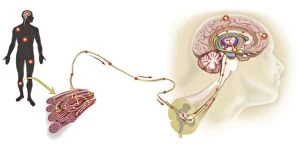
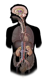
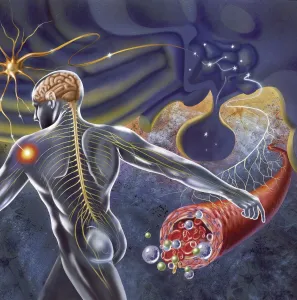
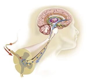
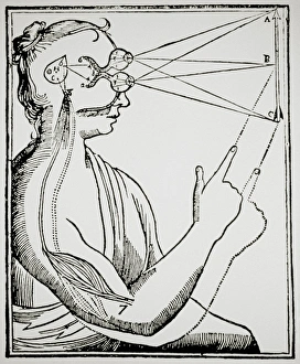
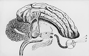
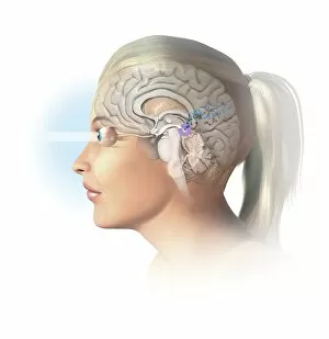
The pineal gland, located in the medulla oblongata of the brain, has long fascinated scientists and philosophers alike. One such philosopher was Rene Descartes, who illustrated the co-ordination of the senses in his artwork from 1692. In his work on vision, Descartes proposed that perceptions travel from the eyes to the pineal gland, which then allows humors to pass to the muscles and produce a response. Descartes' ideas about reflex action and reciprocal innervation were further explored in his book De homine, published in 1662. The annotated title page of this first edition highlights Descartes' belief that the pineal gland is not only responsible for these actions but also serves as the seat of the soul. Biomedical illustrations provide us with a cross-section view of both male and female endocrine systems, showcasing how intricately connected they are to our overall well-being. Additionally, they reveal how our autonomic nervous system and limbic system play vital roles in regulating various bodily functions. A woodcut from Descartes' Treatise of Man depicts his theory that perceptions travel through sensory nerves to reach the pineal gland before transmitting signals to muscles for response production. This illustration emphasizes how crucial this tiny organ is in facilitating communication within our body. Furthermore, we can observe a pathway where pain messages travel via sensory nerves when a muscle is injured. This demonstrates how interconnected our nervous system is with different parts of our body. The sympathetic nervous system's role becomes evident when considering fight-or-flight responses triggered by certain organs during times of stress or danger. It showcases just one aspect of how complexly intertwined our bodies are with their surroundings. Lastly, schematic representations show us how nerve impulses from throughout our body converge on the hypothalamus—a key player involved in maintaining homeostasis—highlighting its significance as an information hub within our central nervous system.