Fraud Blocker Skip to main content

Physiology Collection (page 6)

"Unlocking the Secrets of the Human Body

Background imagePhysiology Collection: Benjamin Ward Richardson, British physician and writer on medical history, 1883

Benjamin Ward Richardson, British physician and writer on medical history, 1883. Artist: Lock & Whitfield
Benjamin Ward Richardson, British physician and writer on medical history, 1883. Richardson was an eminent physician, anaesthetist and physiologist

Background imagePhysiology Collection: William Benjamin Carpenter, CB, MD, LLD, FRS, FLS, FGS, English physiologist and naturalist, 1883

William Benjamin Carpenter, CB, MD, LLD, FRS, FLS, FGS, English physiologist and naturalist, 1883. From Men of Mark: a gallery of contemporary portraits of men distinguished in the Senate

Background imagePhysiology Collection: Hermann Ludwig Ferdinand von Helmholtz, c1900

Hermann Ludwig Ferdinand von Helmholtz, c1900
Hermann Ludwig Ferdinand von Helmholtz, German physicist and physiologist, c1900. The inventor of the opthalmoscope (1850)

Background imagePhysiology Collection: Hermann Ludwig Ferdinand von Helmholtz, German physicist and physiologist, 1907

Hermann Ludwig Ferdinand von Helmholtz, German physicist and physiologist, 1907. The inventor of the opthalmoscope (1850)

Background imagePhysiology Collection: John Scott Burdon-Sanderson, British physiologist, 1894. Artist: Spy

John Scott Burdon-Sanderson, British physiologist, 1894. Artist: Spy
John Scott Burdon-Sanderson, British physiologist, 1894. Burdon-Sanderson (1828-1905) was Regius professor of medicine at Oxford

Background imagePhysiology Collection: Half-title of De Motu Animalum by Giovanni Borelli, 1710

Half-title of De Motu Animalum by Giovanni Borelli, 1710. Giovanni Alfonso Borelli (1608-1679), Italian physiologist and physician, first published this book in 1680-1686

Background imagePhysiology Collection: Harvey demonstrating circulation of the blood to the College of Physicians, c1628 (1870)

Harvey demonstrating circulation of the blood to the College of Physicians, c1628 (1870). William Harvey (1578-1657), English physician, published his famous De motu cordis

Background imagePhysiology Collection: Albrecht von Haller, 18th century Swiss physician and scientist, c1840

Albrecht von Haller, 18th century Swiss physician and scientist, c1840
Albrecht von Haller, 18th century Swiss physician and scientist, [c1840]. Von Haller (1708-1777) worked in the field of experimental physiology and was the founder of the science of neurology

Background imagePhysiology Collection: Albrecht von Haller, Swiss physician and scientist, c1770 (c1780)

Albrecht von Haller, Swiss physician and scientist, c1770 (c1780). Von Haller (1708-1777) worked in the field of experimental physiology and was the founder of the science of neurology

Background imagePhysiology Collection: Rene Descartes diagram of the human brain and eye, 1692

Rene Descartes diagram of the human brain and eye, 1692. From Opera Philosophica by Rene Descartes. (Frankfurt-am-Main, 1692). Originally published in his Tractatus de homine. (Paris, 1664)

Background imagePhysiology Collection: Rene Descartes illustration of the co-ordination of the senses, 1692

Rene Descartes illustration of the co-ordination of the senses, 1692. A visual stimulus travelling from the eye to the pineal gland, H, stops attention being given to an olfactory stimulus

Background imagePhysiology Collection: Rene Descartes idea of vision, showing the function of the eye, optic nerve and brain, 1692

Rene Descartes idea of vision, showing the function of the eye, optic nerve and brain, 1692. From Opera Philosophica by Rene Descartes. (Frankfurt-am-Main, 1692)

Background imagePhysiology Collection: Descartes representation of the antagonistic eye muscles, 1692

Descartes representation of the antagonistic eye muscles, 1692. When E is relaxed A is innervated. Rene Decartes (1596-1650)

Background imagePhysiology Collection: Reverse of medal commemorating 50 years of cinematography by the Lumiere brothers, 1945

Reverse of medal commemorating 50 years of cinematography by the Lumiere brothers, 1945. August (1862-1954) and Louis Lumiere (1864-1948), French chemists and pioneers of cinematography, 1945

Background imagePhysiology Collection: Obverse of medal commemorating 50 years of cinematography by the Lumiere brothers, 1945

Obverse of medal commemorating 50 years of cinematography by the Lumiere brothers, 1945. August (1862-1954) and Louis Lumiere (1864-1948), French chemists and pioneers of cinematography

Background imagePhysiology Collection: Louis Jean Lumiere (1864-1948), pioneer of cinematography, c1935

Louis Jean Lumiere (1864-1948), pioneer of cinematography, c1935
Louis Jean Lumiere (1864-1948), French chemist and pioneer of cinematography, c1935. Collaborated with his elder brother, Auguste. From obverse of commemorative plaquette

Background imagePhysiology Collection: Etienne Jules Marey (1830-1903), French physiologist, 19th century

Etienne Jules Marey (1830-1903), French physiologist, 19th century. Pioneer of cinematography. From reverse of commemorative plaquette showing Marey at his desk

Background imagePhysiology Collection: Etienne Jules Marey (1830-1903), with cine camera, 1903

Etienne Jules Marey (1830-1903), with cine camera, 1903
Etienne Jules Marey (1830-1903), French physiologist, with cine camera, 1903. Top: Chambre chrono-photographique, the first cine-camera, is used to study movement of creatures in aquarium

Background imagePhysiology Collection: Hermann von Helmholtz (1821-1894), German physicist and physiologist, 1894

Hermann von Helmholtz (1821-1894), German physicist and physiologist, 1894
Hermann Helmholtz (1821-1894). German physicist and physiologist, inventor of the Opthalmascope. Seen here giving a lecture on 7 July 1894 shortly before his death

Background imagePhysiology Collection: Descartes illustration of how the distance of an object is perceived with binocular vision, 1692

Descartes illustration of how the distance of an object is perceived with binocular vision, 1692
Descartes (1596-1650) illustration of how the distance of an object is perceived with binocular vision, 1692. From Rene Descartes Opera Philosophica (Tractatus de Homine), 1692

Background imagePhysiology Collection: Luigi Galvanis experiments with electricity, 1791

Luigi Galvanis experiments with electricity, 1791. An electrostatic machine, a Leyden jar and various experiments conducted by Italian physiologist Galvani (1737-1798)

Background imagePhysiology Collection: Descartes explanation of vision, 1692

Descartes explanation of vision, 1692. Light rays being passed through the eye, being focused by the lens (I) and forming images T, S, R on the retina

Background imagePhysiology Collection: Descartes idea of vision, 1692

Descartes idea of vision, 1692
Descartes (1596-1650) idea of vision, [1692]. The passage of nervous impulses from the eye to the pineal gland and so to the muscles. From Rene Descartes Opera Philosophica (Tractatus de homine), 1692

Background imagePhysiology Collection: Descartes idea of how eye passes impulse to brain and so directs a voluntary movement, 1692

Descartes idea of how eye passes impulse to brain and so directs a voluntary movement, 1692. French philosopher and scientist Rene Descartes (1596-1650)

Background imagePhysiology Collection: Involuntary movement, Descartes idea of how impulses from the limbs reach the brain, 1692

Involuntary movement, Descartes idea of how impulses from the limbs reach the brain, 1692. French philosopher and scientist Rene Descartes (1596-1650) believed all nerves to be hollow

Background imagePhysiology Collection: Charles Herbert Best, Canadian physiologist, 1960

Charles Herbert Best, Canadian physiologist, 1960. Bell (1899-1978) with an assistant (left) in the laboratory. Bell assisted Frederick Banting to isolate insulin (1921)

Background imagePhysiology Collection: Circulation of the blood, 1628

Circulation of the blood, 1628. English physician William Harvey (1578-1657) was the first to correctly describe the mechanism whereby blood is circulated in the body

Background imagePhysiology Collection: Gardener smelling a carnation or pink (Dianthus), c1750. Artist: Thomas Kitchin

Gardener smelling a carnation or pink (Dianthus), c1750. Artist: Thomas Kitchin
Gardener smelling a carnation or pink (Dianthus), c1750. French philosopher Rene Descartes interaction theory stated that the reflexes were mechanistically determined and that body

Background imagePhysiology Collection: Diagram of Pavlovs findings on conditioned salivary reflex

Diagram of Pavlovs findings on conditioned salivary reflex. D: organ of touch and temperature; N: 1-6 analysers; S: salivary gland; Q: saliva secreting centre in the medulla oblongata; M: muscle; R

Background imagePhysiology Collection: George von Bekesy (1899-1972), Hungarian-born American physiologist

George von Bekesy (1899-1972), Hungarian-born American physiologist. He won the Nobel prize for physiology or medicine in 1961 for his pioneering research into the function of the inner ear

Background imagePhysiology Collection: Frederick Grant Banting (1891-1941), Canadian physiologist, 1923

Frederick Grant Banting (1891-1941), Canadian physiologist, 1923. Banting and his assistant, CH Best, discovered insulin in 1921

Background imagePhysiology Collection: animal head, animal themes, bird, cross section, long, no people, physiology, profile

animal head, animal themes, bird, cross section, long, no people, physiology, profile, studio shot, tongue, two animals, vertical, watercolour painting, white background, wildlife, woodpecker

Background imagePhysiology Collection: The Muscular System of the Human Body

The Muscular System of the Human Body - from the front and the rear. Date: circa 1790

Background imagePhysiology Collection: SCHWANN, Theodor (1810 - 1882). German physiologist

SCHWANN, Theodor (1810 - 1882). German physiologist, anatomist and naturalist. He developed the cell theory. Oil

Background imagePhysiology Collection: Anterior cross section of spinal column revealing how vertebra fits round spinal cord

Anterior cross section of spinal column revealing how vertebra fits round spinal cord

Background imagePhysiology Collection: Lateral cross section through the skull showing the brain with intermediate layers (meninges)

Lateral cross section through the skull showing the brain with intermediate layers (meninges) highlighted

Background imagePhysiology Collection: Cross section illustration of human ovary

Cross section illustration of human ovary

Background imagePhysiology Collection: Illustration of reptile, bird, rabbit and human embryos in early stage of development

Illustration of reptile, bird, rabbit and human embryos in early stage of development

Background imagePhysiology Collection: Illustration of smell sensors, nasal epithelium, olfactory bulb, turbinate, bones

Illustration of smell sensors, nasal epithelium, olfactory bulb, turbinate, bones, frontal sinus and sphenoid sinus in nasal cavity

Background imagePhysiology Collection: Illustration of cell nucleus containing cell cytoplasm, mitochondria units, DNA, and chromosome

Illustration of cell nucleus containing cell cytoplasm, mitochondria units, DNA, and chromosome

Background imagePhysiology Collection: Digital illustration of penguin circulation showing countercurrent mechanism of warm blood flowing f

Digital illustration of penguin circulation showing countercurrent mechanism of warm blood flowing f

Background imagePhysiology Collection: Cross section digital illustration of Sea Urchin showing gonads, intestine, ampullae and radial cana

Cross section digital illustration of Sea Urchin showing gonads, intestine, ampullae and radial cana

Background imagePhysiology Collection: Digital cross section illustration of male reproductive system

Digital cross section illustration of male reproductive system

Background imagePhysiology Collection: Digital illustration of human breast anatomy, cross section

Digital illustration of human breast anatomy, cross section

Background imagePhysiology Collection: Digital illustration of showing front view of human brain

Digital illustration of showing front view of human brain

Background imagePhysiology Collection: Illustration of anatomy of frog

Illustration of anatomy of frog

Background imagePhysiology Collection: Illustration of lungs and respiratory system of a chimpanzee

Illustration of lungs and respiratory system of a chimpanzee

Background imagePhysiology Collection: Illustration of female human body showing skeleton, brain, heart, lungs, digestive system

Illustration of female human body showing skeleton, brain, heart, lungs, digestive system, and uterus


All Professionally Made to Order for Quick Shipping

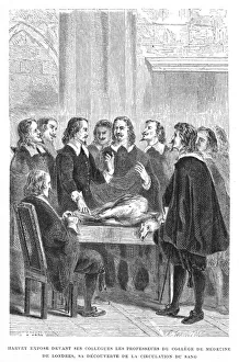
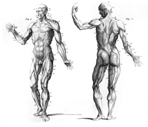
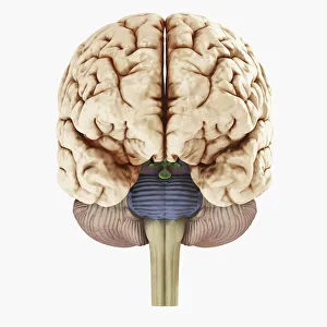
"Unlocking the Secrets of the Human Body: Exploring Physiology from Head to Toe" Dive into the intricate world of human physiology with a captivating view of the anatomy of the brain, seen from an inferior perspective. Step back in time and witness Joseph Wright's masterpiece, "The Airpump, " as it symbolizes our quest to understand how our bodies function on a physiological level. Discover the hidden powerhouses behind every smile and frown with a detailed diagram showcasing facial muscles labeled for your convenience. Take a front-row seat to unravel the complexity of facial muscles as they work harmoniously together, providing us with countless expressions that define our humanity. Delve into knee joint mechanics and gain insight into how this remarkable structure allows us to walk, run, and perform various movements effortlessly. Journey beneath your skin's surface and explore the fascinating relationship between hair follicles and skin – two interconnected systems that contribute to our unique appearance. Witness synapse nerve junctions through a TEM image, offering a glimpse into one of nature's most efficient communication networks within our nervous system. Marvel at the intricacies of head muscles as they enable us to express emotions, chew food, speak articulately, or simply turn our heads towards something captivating. Uncover ancient evolutionary structures within our brains by examining cross-section illustrations depicting limbic system functions alongside primitive forebrain regions. Embark on an enlightening tour through your body's drainage system - discover how lymphatic vessels play an essential role in maintaining overall health and immunity. Explore superior views of colored lobes accompanied by labels; see how different brain regions contribute uniquely to cognition, emotion regulation & sensory processing Dissecting human brain anatomy from lateral view reveals its awe-inspiring complexity; marvel at its convoluted folds & diverse functional areas shaping who we are.