
Phagocytic Collection
Phagocytic cells, such as dendritic cells and macrophages, play a crucial role in our immune system's defense against various pathogens


All Professionally Made to Order for Quick Shipping
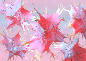
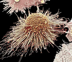
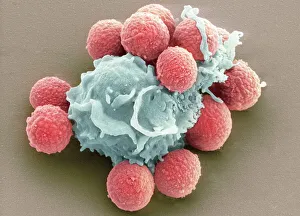
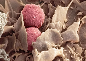
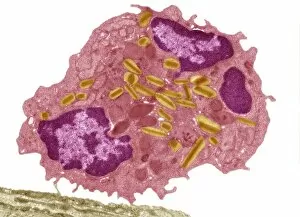
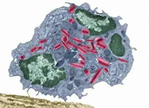
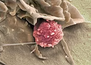
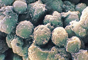
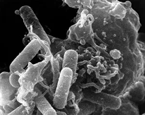
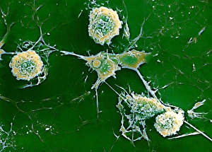
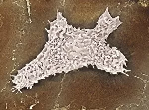
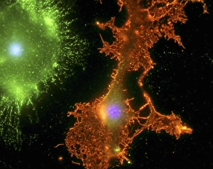
Phagocytic cells, such as dendritic cells and macrophages, play a crucial role in our immune system's defense against various pathogens. This captivating artwork showcases the intricate process of phagocytosis, where these specialized cells engulf and destroy harmful entities. In one mesmerizing image captured by scanning electron microscopy (SEM), we witness the phagocytosis of fungal spores. The magnified view reveals the remarkable ability of these cells to engulf and neutralize potential threats. Another SEM image portrays an activated macrophage in action, showcasing its formidable power to eliminate foreign invaders. These mighty warriors tirelessly patrol our bodies, ensuring that no pathogen goes unnoticed or unchallenged. The microscopic world comes alive through SEM images depicting multiple sclerosis. This debilitating disease affects the central nervous system, highlighting the importance of phagocytes in maintaining its health and integrity. Artistic renditions of dendritic cells further emphasize their critical role in immune responses. Their unique structure allows them to capture antigens efficiently and present them to other immune cells for recognition and activation. Transmission electron microscopy (TEM) provides us with detailed glimpses into cellular processes like phagocytosis. One TEM image captures macrophage cells engrossed in their duty, ready to devour any intruders that dare enter our bodies. Eosinophil white blood cell TEM images showcase their involvement not only in combating infections but also regulating allergic reactions. These specialized phagocytes exhibit extraordinary versatility when it comes to defending our well-being. Through SEM images capturing activated macrophages at work, we witness firsthand their relentless pursuit of protecting us from harm's way. Their robust presence ensures that no invader can escape detection or elimination without consequences. TEM imagery highlights eosinophil white blood cell activity once again—these incredible defenders are always on high alert against potential threats lurking within our bodies' depths. This collection of captivating images and artwork showcases the remarkable world cells.