Fraud Blocker Skip to main content

Parasitism Collection (page 4)

"Unseen Intruders: Exploring the World of Parasitism" Caption: In this captivating series of images, we delve into the intricate and often hidden world of parasitism

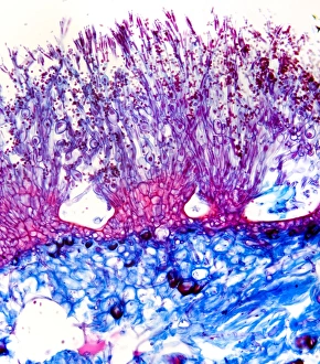
Background imageParasitism Collection: Penicillium mould, light micrograph

Penicillium mould, light micrograph
Penicillium mould. Light micrograph of a section through an orange rind infected with Penicillium sp. blue mould fungus, showing fungal hyphae and conidiophores (pink and purple) with spores (red)

Background imageParasitism Collection: Downy mildew infection, light micrograph

Downy mildew infection, light micrograph
Downy mildew infection. Light micrograph of a section through a grapevine (Vitis sp.) leaf infected with downy mildew (Plasmopara viticola)

Background imageParasitism Collection: Hollyhock rust fungus in a leaf

Hollyhock rust fungus in a leaf. Light micrograph of a section through a hollyhock (Alcea sp.) leaf infected with the parasitic Puccinia malvacearum rust, showing the teliospores (spores)

Background imageParasitism Collection: Clubroot infection, light micrograph

Clubroot infection, light micrograph
Clubroot infection. Light micrograph a section through a cabbage root infected with clubroot (Plasmodiophora brassicae). Plasmodiophora is a parasitic protist that infects the roots of cabbages

Background imageParasitism Collection: Cistanche phelypaea

Cistanche phelypaea flowering on salt marshes in a Algarve, Portugal. This plant is a parasite on Chenopdiaceae

Background imageParasitism Collection: Cytinus hypocistis

Cytinus hypocistis flowering in Algarve, Portugal. This is an ant pollinated root parasite of Cistus species

Background imageParasitism Collection: Naked Broomrape (Orobanche uniflora)

Naked Broomrape (Orobanche uniflora), also known as One-flowered Broomrape, flowering in Waterton, Canada. This plant is parasitic on Round-leaved Alumroot

Background imageParasitism Collection: Monotropa hypopitys flowers

Monotropa hypopitys flowers
Monotropa hypopitys district Närke

Background imageParasitism Collection: Bumblebee drinking

Bumblebee drinking. Buff-tailed bumblebee (Bombus terrestris) drinking rainwater off a leaf. An orange mite is visible on its thorax. Photographed in Italy

Background imageParasitism Collection: Cottony cushion scale bugs

Cottony cushion scale bugs. Cluster of cottony cushion scale (Icerya purchasi) bugs and their egg cases on a lemon tree. Photographed in Italy

Background imageParasitism Collection: Edoardo Perroncito, Italian physician

Edoardo Perroncito, Italian physician
Edoardo Perroncito (1847Ôé¼" 1936), Italian physician, veterinarian and parasitologist. Perroncito is credited, along with Luigi Pagliani (1847Ôé¼" 1931)

Background imageParasitism Collection: Human parasites, historical artwork

Human parasites, historical artwork
Human parasites. 1575 artwork of human parasites drawn by the Dutch physician, astronomer and astrologer Cornelius Gemma (1535Ôé¼" 1579)

Background imageParasitism Collection: Giovanni Grassi, Italian zoologist

Giovanni Grassi, Italian zoologist
Giovanni Grassi. Portrait of Giovanni Batista Grassi (1854-1925), Italian zoologist and malaria pioneer. In 1889-90 Grassi suggested that different species of the Plasmodium protozoa caused different

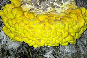
Background imageParasitism Collection: Bracket fungus (Laetiporus sulphureus)

Bracket fungus (Laetiporus sulphureus)
Bracket fungus. Young sulphur-coloured fruiting body of the edible bracket fungus known as the chicken of the woods (Laetiporus sulphureus), growing on a living willow tree

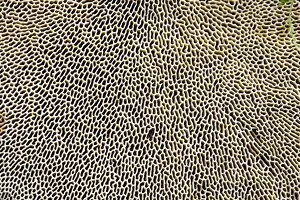
Background imageParasitism Collection: Bracket fungus (Daedaleopsis confragosa)

Bracket fungus (Daedaleopsis confragosa)
Blushing bracket fungus (Daedaleopsis confragosa). Close-up of the underside of the blushing bracket fungus showing its pores. The pores are the ends of the tubes within which the spores are produced

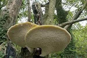
Background imageParasitism Collection: Bracket fungus (Polyporus squamosus)

Bracket fungus (Polyporus squamosus)
Bracket fungus. Dryads saddle (Polyporus squamosus) bracket fungus on a tree

Background imageParasitism Collection: Artichoke Galls on Pedunculate Oak

Artichoke Galls on Pedunculate Oak (Quercus rubus). Flower buds infected by the gall wasp Andricus fecundator. In spring the female gall wasp lays single eggs in buds of the Pedunculate oak

Background imageParasitism Collection: Chrysalis infected with parasitic larvae

Chrysalis infected with parasitic larvae. Chrysalis of the small tortoiseshell butterfly (Aglais urticae) with a thin thread extending from it

Background imageParasitism Collection: Dodder parasitic plant

Dodder parasitic plant. Polarised light micrograph of a section through a host plant parasitised by a dodder (Cuscuta sp.)

Background imageParasitism Collection: Plant parasite, light micrograph

Plant parasite, light micrograph
Plant parasite. Polarised light micrograph of a transverse section through a plant stem (left) entwined by the common dodder (Cuscuta sp.) parasitic plant (round, right)

Background imageParasitism Collection: Chrysalise infected with parasitic larvae

Chrysalise infected with parasitic larvae
Chrysalises infected with parasitic larvae. Chrysalises of the small tortoiseshell butterfly (Aglais urticae) with thin threads extending from them

Background imageParasitism Collection: Cardinalfish and parasite

Cardinalfish and parasite. Cardinalfish (family Apogonidae) with a parasitic isopod crustacean on its head. Parasitic isopods feed on the tissues of their host

Background imageParasitism Collection: Nettle leaf gall midge larva

Nettle leaf gall midge larva, artwork. Cross- section through a stinging nettle leaf (Urtica dioica) and leaf gall. The gall is inhabited by the parasitic larva of the nettle gall midge

Background imageParasitism Collection: Tooth fungi (Hericium coralloides)

Tooth fungi (Hericium coralloides) on a host tree. The mushroom of this fungi bears its spores on a hymenium (a layer of mother cells)

Background imageParasitism Collection: Nettle leaf galls

Nettle leaf galls, artwork. Four views of galls at the base of a stinging nettle leaf (Urtica dioica). These abnormal outgrowths are caused by the parasitic larvae of the nettle gall midge

Background imageParasitism Collection: Desert dodder (Cuscuta denticulata)

Desert dodder (Cuscuta denticulata)
Desert dodder (Cuscuta denticulata, orange) parasitising a white burro bush plant (Ambrosia dumosa). Photographed in the USA

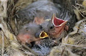
Background imageParasitism Collection: Bronzed Cowbird, Molothrus aeneus, young in nest of Bewicks Wren, Brood Parasit

Bronzed Cowbird, Molothrus aeneus, young in nest of Bewicks Wren, Brood Parasit, Starr County, Rio Grande Valley, Texas, USA

Background imageParasitism Collection: Mistletoe Viscum album with berries, midwinter. Widespread semi-parasite of trees. On apple, Dorset

Mistletoe Viscum album with berries, midwinter. Widespread semi-parasite of trees. On apple, Dorset
ROG-13360 Mistletoe - with berries, midwinter Widespread semi-parasite of trees. On apple, Dorset. UK Viscum album Bob Gibbons Please note that prints are for personal display purposes only

Background imageParasitism Collection: Parasitic Tachinid fly - on Angelica; parasite on lepidoptera larvae

Parasitic Tachinid fly - on Angelica; parasite on lepidoptera larvae
ROG-14099 Parasitic Tachinid fly - on Angelica; parasite on lepidoptera Tachina fera Bob Gibbons Please note that prints are for personal display purposes only and may not be reproduced in any way

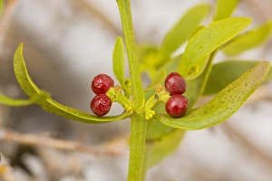
Background imageParasitism Collection: Red-berried Mistletoe - in hawthorn high in the Middle Atlas Mountains, Morocco

Red-berried Mistletoe - in hawthorn high in the Middle Atlas Mountains, Morocco
ROG-13578 Red-berried Mistletoe - in hawthorn high in the Middle Atlas Mountains Morocco. Viscum cruciatum Bob Gibbons Please note that prints are for personal display purposes only

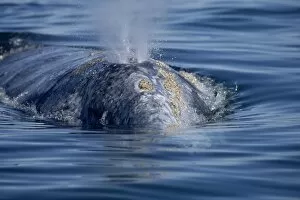
Background imageParasitism Collection: Grey whale (Eschrichtius robustus) surfacing to blow near boat. Yellowish barnacles

Grey whale (Eschrichtius robustus) surfacing to blow near boat. Yellowish barnacles
Tom Walmsley / SplashdownDirect


All Professionally Made to Order for Quick Shipping

"Unseen Intruders: Exploring the World of Parasitism" Caption: In this captivating series of images, we delve into the intricate and often hidden world of parasitism. Starting with a close-up view captured by Scanning Electron Microscopy (SEM), we witness the astonishing presence of eyelash mites (Picture No. 12479415). These tiny creatures, barely visible to the naked eye, inhabit our very own eyelashes, reminding us that even within our bodies, parasites can thrive. Zooming in further with SEM technology, another image reveals the intriguing tails of these eyelash mites (Picture No. 12479414). Their peculiar structures hint at their evolutionary adaptations for survival and reproduction. Moving beyond microscopic organisms, we encounter an extraordinary example of brood parasitism involving a Cuckoo chick in a Reed Warbler nest (Acrocephalus scirpaceus) (INDO 20-06 393). Picture No. 12479506 showcases a twelve-day-old European Cuckoo chick nestled among unsuspecting Reed Warblers in Africa. This remarkable behavior is not without consequences as native species are affected by sleeping sickness due to such intrusions. The historical engraving by Karl Rudolphi portrays this phenomenon vividly; it serves as a reminder that parasitic interactions have fascinated naturalists throughout history (Picture No. 12479416). The image depicts an African scene where a twelve-day-old European Cuckoo chick thrives at the expense of its foster parents' offspring while contributing to ecological imbalances. Lastly, we witness firsthand how nature's delicate balance can be disrupted when observing a Reed Warbler diligently feeding its adopted twelve-day-old Cuckoo chick (Cuculus canorus) despite being unrelated species (Acrocephalus scirpaceus). These captivating visuals shed light on the intricacies and complexities surrounding parasitism—a phenomenon both fascinating and often unsettling.