Fraud Blocker Skip to main content

Oesophageal Collection

The oesophagus, also known as the food pipe, plays a vital role in our digestive system

Background imageOesophageal Collection: Throat cancer, X-rays

Throat cancer, X-rays
Throat cancer. Coloured X-rays showing the transit of a contrast medium (dense white) through the throat of a 54-year-old patient with an adenocarcinoma (cancer) of the lower oesophagus (gullet)

Background imageOesophageal Collection: Hiatal hernia, endoscopic view C016 / 8330

Hiatal hernia, endoscopic view C016 / 8330
Hiatal hernia. Endoscopic view of a hiatal hernia (gastro-oesophageal prolapse), seen from below, with the endoscope(black) seen in an inverted position inside the stomach

Background imageOesophageal Collection: Hiatal hernia, endoscopic view C016 / 8329

Hiatal hernia, endoscopic view C016 / 8329
Hiatal hernia. Endoscopic view of a hiatal hernia (gastro-oesophageal prolapse), seen from above. A hernia is the protrusion of an organ, or the fascia of an organ

Background imageOesophageal Collection: Oesophagus wall, light micrograph

Oesophagus wall, light micrograph
Oesophagus wall. Coloured light micrograph of a section through the human oesophagus, which passes food from the mouth to the stomach

Background imageOesophageal Collection: Oesophagus, SEM

Oesophagus, SEM
Oesophagus. Coloured scanning electron micrograph (SEM) of a freeze-fracture through the muscularis externa of the oesophagus. This forms part of the muscularis mucosa found throughout the gut

Background imageOesophageal Collection: Oesophagus epithelium, SEM

Oesophagus epithelium, SEM
Oesophagus epithelium. Coloured scanning electron micrograph (SEM) of the lining of the human oesophagus or gullet (yellow) and two unknown bacteria (pink & purple)

Background imageOesophageal Collection: Oesophagus lining, SEM

Oesophagus lining, SEM
Oesophagus lining. Coloured scanning electron micrograph (SEM) of the lining (epithelium) of the human oesophagus (gullet, blue) and two bacteria (red)

Background imageOesophageal Collection: Swallowed coin, X-ray

Swallowed coin, X-ray
Swallowed coin. Coloured X-ray of a coin (orange) in a childs oesophagus. The heart (blue, centre), ribcage (upper centre) and abdominal region (blue, lower centre) are all seen

Background imageOesophageal Collection: Mouth and throat anatomy, artwork

Mouth and throat anatomy, artwork


All Professionally Made to Order for Quick Shipping

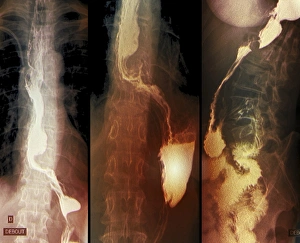
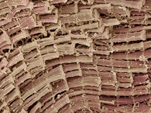
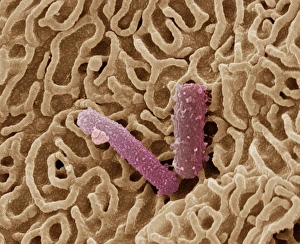
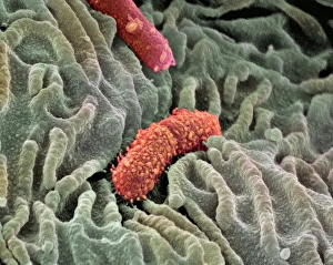
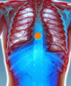
The oesophagus, also known as the food pipe, plays a vital role in our digestive system. However, it is not immune to certain health conditions that can affect its function. One such condition is throat cancer, which can be detected through X-rays. These images help medical professionals identify any abnormalities or tumors within the oesophagus. Examining the oesophagus under a light micrograph reveals intricate details of its wall structure. This microscopic view allows scientists to study the composition and organization of cells within the oesophageal wall. Similarly, an endoscopic view provides valuable insights into conditions like hiatal hernia, where part of the stomach protrudes into the chest cavity through an opening in the diaphragm. To further understand the oesophagus at a cellular level, scanning electron microscopy (SEM) offers detailed imagery of its lining and epithelium. SEM images showcase the delicate yet resilient nature of these tissues that protect and facilitate smooth passage for food during digestion. By combining various diagnostic techniques such as X-rays and microscopic examinations, healthcare professionals gain a comprehensive understanding of any potential issues affecting this crucial organ. Early detection and accurate diagnosis are essential in managing diseases related to the oesophagus effectively. Exploring different aspects of "oesophageal" health using advanced imaging technologies like X-rays and SEM aids in identifying conditions such as throat cancer or hiatal hernia while shedding light on its complex structure at both macroscopic and microscopic levels.