Fraud Blocker Skip to main content

Nuclei Collection (page 3)

"Nuclei: The Intricate Powerhouses within Our Cells and Beyond" Delving into the microscopic world, we encounter the mesmerizing beauty of nuclei

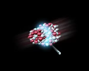
Background imageNuclei Collection: Heavy atomic nuclei colliding, artwork

Heavy atomic nuclei colliding, artwork
Heavy atomic nuclei colliding. Computer artwork of two heavy atomic nuclei colliding. The collision has created a subatomic particle (lower right)

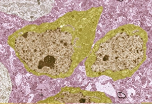
Background imageNuclei Collection: Brain cells, TEM C013 / 4801

Brain cells, TEM C013 / 4801
Brain cells. Transmission electron micrograph (TEM) of a section through oligodendrocytes (dark) in human brain tissue, showing free ribosomes (dark green dots), golgi apparatus (curved brown lines)

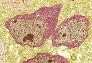
Background imageNuclei Collection: Brain cells, TEM C013 / 4800

Brain cells, TEM C013 / 4800
Brain cells. Transmission electron micrograph (TEM) of a section through oligodendrocytes in human brain tissue, showing free ribosomes (dark pink dots), golgi apparatus (curved brown lines)

Background imageNuclei Collection: Brain cell, TEM C013 / 4799

Brain cell, TEM C013 / 4799
Brain cell. Transmission electron micrograph (TEM) of a section through an oligodendrocyte in human brain tissue, showing free ribosomes (dark brown dots), golgi apparatus (curved orange lines)

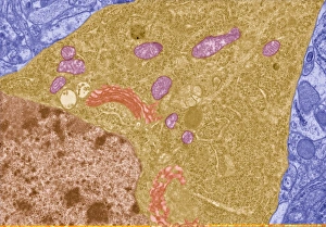
Background imageNuclei Collection: Brain cell, TEM C013 / 4798

Brain cell, TEM C013 / 4798
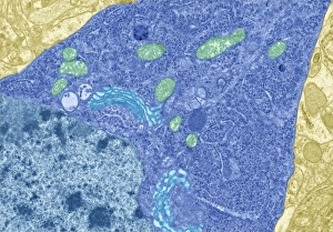
Brain cell. Transmission electron micrograph (TEM) of a section through an oligodendrocyte in human brain tissue, showing free ribosomes (dark blue dots), golgi apparatus (curved light blue lines)

Background imageNuclei Collection: HeLa cells, light micrograph C013 / 4774

HeLa cells, light micrograph C013 / 4774
HeLa cells. Multi-photon fluorescence light micrograph of a group of cultured HeLa cells, showing the cell nuclei, which contain the cells genetic information (DNA, red)

Background imageNuclei Collection: HeLa cells, light micrograph C013 / 4773

HeLa cells, light micrograph C013 / 4773
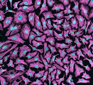
HeLa cells. Multi-photon fluorescence light micrograph of a group of cultured HeLa cells, showing the cell nuclei, which contain the cells genetic information (DNA, blue), and microtubules (pink)

Background imageNuclei Collection: Giardia protozoan, TEM

Giardia protozoan, TEM
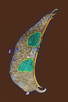
Giardia protozoan. Coloured transmission electron micrograph (TEM) of a Giardia sp. protozoan. It is a single-celled organism that has two cell nuclei (green)

Background imageNuclei Collection: Naegleria fowleri protozoa, TEM

Naegleria fowleri protozoa, TEM
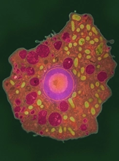
Naegleria fowleri protozoan. Coloured transmission electron micrograph (TEM) of a section through a Naegleria fowleri protozoan

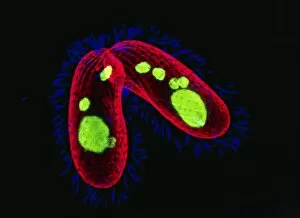
Background imageNuclei Collection: Tetrahymena protozoa

Tetrahymena protozoa. Immunofluorescent light micrograph of two Tetrahymena thermophila protozoa (single-celled animals). Nuclei are green, cell walls red and cilia (hairs) blue. T

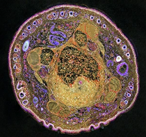
Background imageNuclei Collection: Mouse tail, light micrograph

Mouse tail, light micrograph
Mouse tail. Light micrograph of a cross-section through a mouses tail. At centre is the tail vein, which contains red blood cells. A smaller vein (purple squiggle) is seen at top centre left

Background imageNuclei Collection: Hydrogen fuel cell, artwork

Hydrogen fuel cell, artwork
Hydrogen fuel cell, computer artwork. This is a clean and efficient power source. Hydrogen is liberated from a natural source such as methanol or natural gases

Background imageNuclei Collection: Embryonic stem cells

Embryonic stem cells. Computer-enhanced confocal light micrograph of dividing stem cells from the ventricular zone of the retina of a developing embryo

Background imageNuclei Collection: Dividing cell, light micrograph

Dividing cell, light micrograph
Cell division. Fluorescent light micrograph of a cell that has divided by mitosis, the asexual replication of a cell into two new cells

Background imageNuclei Collection: Skin cells, TEM

Skin cells, TEM
Skin cells. Coloured transmission electron micrograph (TEM) of a section through keratinocyte skin cells, which are found in the epidermis layer of the skin

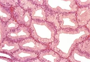
Background imageNuclei Collection: Lacrimal gland, light micrograph

Lacrimal gland, light micrograph
Lacrimal gland. Light micrograph of a section through a lacrimal gland. The lacrimal glands, which are situated one above each eye, secrete tears

Background imageNuclei Collection: Pituitary gland, TEM

Pituitary gland, TEM
Pituitary gland. Coloured transmission electron micrograph (TEM) of cells in the anterior pituitary gland, a hormone-secreting gland at the base of the brain

Background imageNuclei Collection: Kidney tubule, TEM

Kidney tubule, TEM
Kidney tubule. Coloured transmission electron micrograph (TEM) of a section through a proximal convoluted tubule in the kidney

Background imageNuclei Collection: Unstressed cells

Unstressed cells (Image 1 of 2). Immunofluorescent light micrograph of unstressed kidney cells. The nuclei contain the RNA (ribonucleic acid)-binding protein TIA (blue) and DNA (deoxyribonucleic acid)

Background imageNuclei Collection: Kidney tubules in section

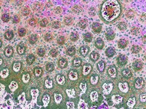
Kidney tubules in section
Kidney tubules. Fluorescent light micrograph of a section through kidney tissue showing numerous tubules (red/green). The tubules are seen in cross-section

Background imageNuclei Collection: Liver cells, light micrograph

Liver cells, light micrograph
Liver cells. Light micrograph of a section through a human liver. The liver cells are pink with purple nuclei. A duct canal and its surrounding tissue can be seen at lower left

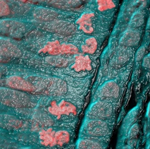
Background imageNuclei Collection: Intestinal cells, light micrograph

Intestinal cells, light micrograph
Intestinal cells. Light micrograph of a section through two folds (villi) in the small intestine. The villi are aligned vertically, with one at left and one at right
Background imageNuclei Collection: Rat tongue, light micrograph

Rat tongue, light micrograph
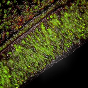
Rat tongue. Confocal light micrograph of an en face section through a rats tongue, showing tastebuds. Actin protein filaments, which make up part of the cytoskeleton, are orange
Background imageNuclei Collection: Secretory cells in pancreas, SEM

Secretory cells in pancreas, SEM
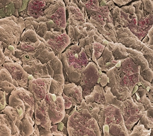
Pancreatic secretory cells. Coloured scanning electron micrograph (SEM) of a freeze-fracture through a healthy pancreas, showing the secretory tissue

Background imageNuclei Collection: Stressed cells

Stressed cells (image 2 of 2). Immunofluorescent light micrograph of stressed kidney cells. Stress is caused by chemicals, UV light, viral infection and heat shock. The cell enters an emergency mode

Background imageNuclei Collection: Lung cells, light micrograph

Lung cells, light micrograph
Lung cells. Light micrograph of a section through the wall of a bronchus in the lungs. This is the upper layer of cells (the epithelium). The cell nuclei (round, dark) are across bottom

Background imageNuclei Collection: Brain cortex tissue, light micrograph

Brain cortex tissue, light micrograph
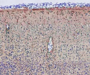
Brain cortex tissue. Light micrograph of a section through tissue from the cortex of the brain. The cortex, or grey matter, is the outer layer of the brain

Background imageNuclei Collection: Gut muscle cells, TEM

Gut muscle cells, TEM
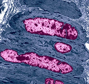
Gut muscle cells, coloured transmission electron micrograph (TEM). Nuclei are pink. The involuntary contraction of this smooth muscle is responsible for peristalsis

Background imageNuclei Collection: Dendritic cell, TEM

Dendritic cell, TEM
Dendritic cell. Coloured transmission electron micrograph (TEM) of a section through dendritic cells. These are part of the human immune system

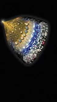
Background imageNuclei Collection: Early history of the universe, artwork

Early history of the universe, artwork
Early history of the universe. Artwork showing the cooling and expansion of the early universe from its origin in the Big Bang (upper left)

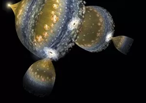
Background imageNuclei Collection: Multiple universes, artwork

Multiple universes, artwork
Multiple universes. Artwork showing multiple universes forming from black holes following the Big Bang formation of the initial universe at top left

Background imageNuclei Collection: Colliding beam fusion reactor

Colliding beam fusion reactor, computer artwork. This is one design for a nuclear fusion reactor that could be used for producing power

Background imageNuclei Collection: Uterus cells, TEM

Uterus cells, TEM
Uterus cells. Coloured transmission electron micrograph (TEM) of endometrial cells (yellow) from the uterus. These cells form the endometrium, the epithelial tissue lining inside the uterus

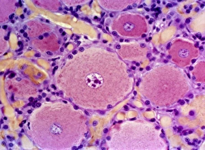
Background imageNuclei Collection: Spinal nerve ganglion, light micrograph

Spinal nerve ganglion, light micrograph
Spinal nerve ganglion. Light micrograph of a cross-section through a spinal nerve ganglion. This is a node of nerve cells located just outside the spinal cord at the point where it is joined by

Background imageNuclei Collection: Neurosphere culture

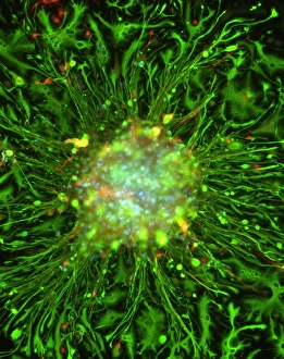
Neurosphere culture. Fluorescent light microscope of a group of neural stem cells (neurosphere) in culture. The stem cells are differentiating into neurons (red) and nerve support cells (green)

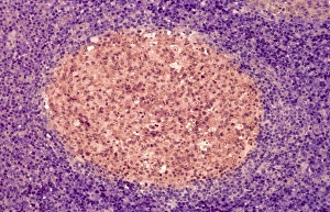
Background imageNuclei Collection: Lymph node, light micrograph

Lymph node, light micrograph
Lymph node. Coloured light micrograph of a section through a lymph node. A lymph node filters pathogens from lymph fluid, a watery liquid that surrounds the tissues of the body

Background imageNuclei Collection: Human blood cells, light micrograph

Human blood cells, light micrograph
Human blood cells. Light micrograph of blood cells at a site of inflammation, showing red blood cells (erythrocytes, red) surrounded by many granulocytes (blue)

Background imageNuclei Collection: Cells

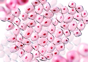
Cells. Computer artwork of non-specific cells. The red dot in the centre of each cell is the nucleus where the DNA (deoxyribonucleic acid) of each cell is stored

Background imageNuclei Collection: HaCaT culture cells

HaCaT culture cells
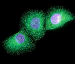
HaCaT cells. Immunofluorescence light micrograph of three HaCaT cells. Their nuclei, which contain the cells genetic information in the form of deoxyribonucleic acid (DNA), are blue

Background imageNuclei Collection: Cell death

Cell death. Computer-enhanced confocal light micrograph of cells in the retina of the eye undergoing programmed cell death (apoptosis)

Background imageNuclei Collection: Cells, SEM

Cells, SEM
Cell cytoskeleton. Coloured scanning electron micrograph (SEM) of the cytoskeleton (blue) and nuclei (green) of cultured cells

Background imageNuclei Collection: Mitosis, artwork

Mitosis, artwork

Background imageNuclei Collection: Smooth muscle, light micrograph

Smooth muscle, light micrograph
Smooth muscle. Light micrograph of a longitudinal section through smooth muscle from the gut. The cells have typically elongated nuclei. Magnification: x240 when printed at 10 centimetres wide


All Professionally Made to Order for Quick Shipping

"Nuclei: The Intricate Powerhouses within Our Cells and Beyond" Delving into the microscopic world, we encounter the mesmerizing beauty of nuclei. In cerebellum tissue, a light micrograph reveals these vital command centers orchestrating our every move. Venturing deeper into scientific frontiers, we find ourselves at CERN's ATLAS detector, where nuclei play a crucial role in unraveling the mysteries of particle physics. Similarly, the CMS detector at CERN unveils their significance in understanding fundamental particles and forces. Shifting our focus to brain anatomy, hippocampus tissue showcases intricate nuclei that contribute to memory formation and spatial navigation. Meanwhile, HeLa cells captured under a light microscope exhibit their own unique nuclei patterns (C017 / 8299), highlighting their importance in medical research. Art meets science as an artwork depicting medulla oblongata reminds us of its critical role in regulating essential bodily functions through its specialized nuclei arrangement. Nuclear fission artwork further emphasizes how they are release immense energy when harnessed correctly. Zooming out to kidney tubules sectioned under a microscope slide unravels the presence of numerous cell nuclei responsible for maintaining fluid balance and waste elimination within our bodies. Expanding beyond traditional boundaries, glial stem cell culture offers insights into regenerative medicine with its vibrant display of proliferating nuclei (light micrograph). These versatile structures are central to cellular growth and differentiation processes. Examining cell structure more broadly uncovers how each nucleus houses genetic material that directs cellular activities like protein synthesis and DNA replication, and is this blueprint that shapes life itself. Lastly, exploring brain tissue blood supply highlights how oxygen-rich blood nourishes countless neuronal networks residing within diverse nuclear ensembles (HeLa cells - C017 / 8298). Intricate yet awe-inspiring, these glimpses into various realms remind us of the indispensable roles played by nucleic entities – from individual cells to complex systems – in shaping our understanding of life, physics, and medicine.