Fraud Blocker Skip to main content

Neurones Collection (page 6)

Neurons, the building blocks of our nervous system, play a crucial role in transmitting information throughout our bodies

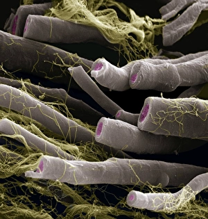
Background imageNeurones Collection: Nerve fibres, SEM

Nerve fibres, SEM
Myelinated nerve fibres, coloured scanning electron micrograph (SEM). The myelin sheath is grey, the axoplasm pink and the endoneurium (connective tissue) yellow

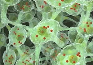
Background imageNeurones Collection: Parkinsons disease nerve cells, artwork

Parkinsons disease nerve cells, artwork
Parkinsons disease nerve cells. Computer artwork of human nerve cells (neurons, green) affected by Lewy bodies (orange) in the brain of someone with Parkinsons disease

Background imageNeurones Collection: Spinal cord, SEM

Spinal cord, SEM
Spinal cord. Coloured scanning electron micrograph (SEM) of a section through a spinal cord, showing the grey matter (green) and white matter (blue)

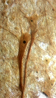
Background imageNeurones Collection: Pyramidal neurons, light micrograph

Pyramidal neurons, light micrograph with Nissl staining. This sample, from a human brain, shows two pyramidal neurons. Pyramidal neurons (nerve cells)

Background imageNeurones Collection: Optic nerve fibres, SEM

Optic nerve fibres, SEM
Optic nerve fibres. Coloured scanning electron micrograph (SEM) of axons from the optic nerve. Connective tissue fibres (thread-like) are also visible

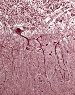
Background imageNeurones Collection: Purkinje neurons, light micrograph

Purkinje neurons, light micrograph

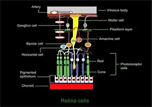
Background imageNeurones Collection: Retina structure

Retina structure
Retinal structure, artwork. Light falling on the retina passes from top to bottom. At bottom is the choroid layer (red), which lines the inside of the eye

Background imageNeurones Collection: Retina blood vessels and nerve cells

Retina blood vessels and nerve cells. Light micrograph showing the blood vessels and nerve cells in the retina of an eye. The retina is the light-sensitive membrane that lines the back of the eyeball

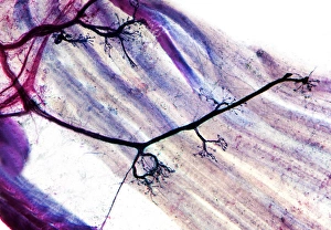
Background imageNeurones Collection: Muscle motor neurones, light micrograph

Muscle motor neurones, light micrograph
Muscle motor neurones. Light micrograph of dendrites (axons, black) from motor nerve cells (motor neurones) in muscle tissue (pink-purple strands)

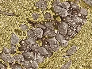
Background imageNeurones Collection: Hippocampal neurons, SEM

Hippocampal neurons, SEM
Hippocampal neurons. Coloured scanning electron micrograph (SEM) of neurons (nerve cells, brown) in the hippocampus of the brain

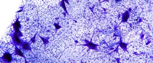
Background imageNeurones Collection: Motor neurones, light micrograph

Motor neurones, light micrograph
Motor neurones. Light micrograph of motor nerve cells (motor neurones) and their support cells. The cell bodies of the neurones (large deep purple areas) have a central nucleus

Background imageNeurones Collection: Nerve synapse, artwork

Nerve synapse, artwork
Nerve synapse. Computer artwork of of a junction, or synapse, between two nerve cells (neurons). As the electrical signal reaches the presynaptic end of a neuron it triggers the release of

Background imageNeurones Collection: Nerve fibre, artwork

Nerve fibre, artwork
Nerve fibre, computer artwork

Background imageNeurones Collection: Embryonic stem cells, light micrograph

Embryonic stem cells, light micrograph
Embryonic stem cells. Fluorescence light micrograph of human embryonic neural stem cells forming neuronal networks. Tubulin protein is red; cell nuclei are blue

Background imageNeurones Collection: DNA and MECP2 complex, molecular mode

DNA and MECP2 complex, molecular mode
DNA and MECP2 complex. Computer artwork showing the molecular structure of MECP2 (methyl CpG binding protein 2 (Rett syndrome)) bound to the BDNF (brain-derived neurotrophic factor)

Background imageNeurones Collection: Neural stem cells, light micrograph

Neural stem cells, light micrograph
Neural stem cells. Light micrograph of human adult neural stem cells grown in suspension as neurospheres. Magnification: x5 when printed 10 centimetres wide

Background imageNeurones Collection: DNA and BDNF gene, molecular model

DNA and BDNF gene, molecular model
DNA and BDNF gene. Molecular model of the methylated BDNF (brain-derived neurotrophic factor) gene (yellow and red) on a strand of DNA (deoxyribonucleic acid)

Background imageNeurones Collection: Synapse, artwork

Synapse, artwork
Synapse. Computer artwork of a synapse, the junction between nerve cells. Synapses transmit electrical signals from one nerve cell to the next

Background imageNeurones Collection: Alzheimers disease, computer artwork

Alzheimers disease, computer artwork
3D computer artwork depicting senile amyloid plaque (brown) in the brain, which kill surrounding neurons (blue). Large numbers of senile plaques are characteristic features of Alzheimers disease

Background imageNeurones Collection: Parkinsons disease, artwork

Parkinsons disease, artwork
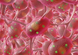
Parkinsons disease. Computer artwork of neurons (nerve cells, pink) containing Lewy bodies (green). Lewy bodies, which are deposits of protein

Background imageNeurones Collection: Taste bud anatomy, diagram

Taste bud anatomy, diagram
Taste bud anatomy. Diagram of the anatomical structure of a taste bud on a tongue. The surface of the tongue is at bottom, with the tastebud (rounded structure) at centre

Background imageNeurones Collection: Neural network, artwork

Neural network, artwork
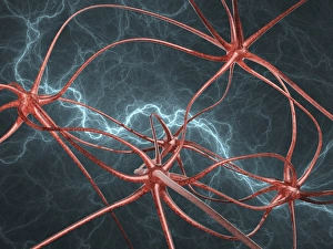
Neural network, computer artwork

Background imageNeurones Collection: Neural network

Neural network. Artwork of nerve cells (neurons, green) connected by nerve processes (dendrites and axons) to form a neural network

Background imageNeurones Collection: Mirror neuron, conceptual image

Mirror neuron, conceptual image
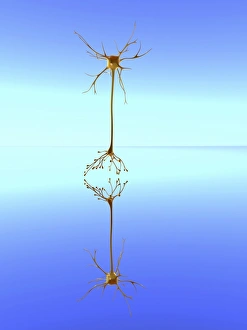
Mirror neuron, conceptual computer artwork. Nerve cell, or neuron, reflected in water, representing a mirror neuron cell. Mirror neurons are found in the brain

Background imageNeurones Collection: Olfactory epithelium, artwork

Olfactory epithelium, artwork
Olfactory epithelium. Computer artwork showing the structure of the specialised layer of tissue that lines the inside of the nasal cavity and is involved in smell

Background imageNeurones Collection: Nerve cell synapse, computer artwork

Nerve cell synapse, computer artwork. Nerve cells, or neurons, are responsible for passing information around the central nervous system (CNS) and from the CNS to the rest of the body

Background imageNeurones Collection: Neuromuscular junctions, artwork

Neuromuscular junctions, artwork
Neuromuscular junctions. Computer artwork of junctions between nerves (thread-like objects) and a muscle (purple surface)

Background imageNeurones Collection: Synapses, artwork

Synapses, artwork
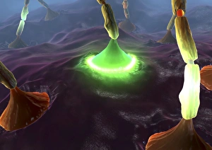
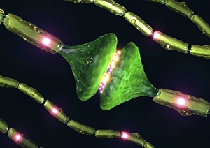
Synapses. Computer artwork of synapses, the junctions between the ends (green, swollen) of two nerve cells (neurons). Signals are passed along nerve cells in the form of an electrical impulse

Background imageNeurones Collection: Glial stem cell culture, light micrograph

Glial stem cell culture, light micrograph
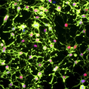
Glial stem cell culture. Fluorescent light micrograph of glial stem cells producing the proteins NG2 (green) and OLIG2 (oligodendrocyte lineage transcription factor 2, red) as they mature

Background imageNeurones Collection: Nerve cell trauma response

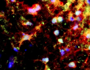
Nerve cell trauma response. Fluorescent light micrograph of a section through a spinal cord affected by multiple sclerosis (MS)

Background imageNeurones Collection: Neural stem cells in culture

Neural stem cells in culture
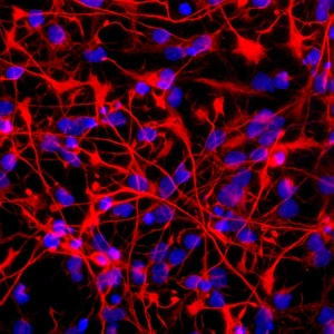
Neural stem cell in culture, fluorescent light micrograph. The stem cells have been dyed for nestin (red), an intermediate filament (IF) protein, and the nuclei are dyed blue

Background imageNeurones Collection: Neural network, abstract artwork

Neural network, abstract artwork
Neural network. Abstract computer artwork representing nerve cells, or neurons. Neurons are responsible for passing information around the central nervous system (CNS)

Background imageNeurones Collection: Neural network, computer artwork

Neural network, computer artwork
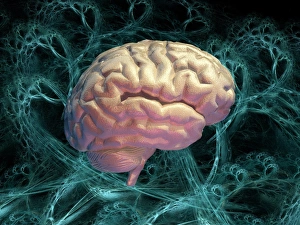
Neural network. Conceptual computer artwork of a brain, with the brains neural network represented by blue strands in the background. The front of the brain is at right


All Professionally Made to Order for Quick Shipping

Neurons, the building blocks of our nervous system, play a crucial role in transmitting information throughout our bodies. These remarkable cells come in various shapes and sizes, each with its own unique function. In a mesmerizing light micrograph, nerve and glial cells are intricately intertwined like a complex network. The delicate synapse nerve junctions captured by transmission electron microscopy (TEM) reveal the microscopic connections that allow neurons to communicate with one another. Glial stem cell culture, seen through another light micrograph, showcases the incredible regenerative potential of these unsung heroes. They possess the ability to differentiate into different types of glial cells which support and protect neurons. Examining brain tissue blood supply under the microscope reveals an intricate web of vessels nourishing this vital organ. This lifeline ensures that neurons receive oxygen and nutrients necessary for their proper functioning. Another captivating image shows neural stem cell culture - a glimpse into the fascinating world of neurogenesis where new neurons are born. This process holds immense promise for understanding brain development and treating neurological disorders. Zooming closer into cerebral cortex nerve cells brings forth their intricate structure and complexity. These specialized neurons enable us to think, reason, learn, and perceive the world around us. Even tiny creatures like C. Elegans worms have their neuronal beauty revealed through light microscopy; they serve as valuable models for studying fundamental aspects of neuroscience research. Motor neurons depicted in yet another stunning light micrograph remind us how essential they are for coordinating movement throughout our body - from simple reflexes to complex actions requiring precision control. Transmission electron microscopy (TEM) offers an up-close look at individual nerve cells' ultrastructure: revealing their organelles such as mitochondria or Golgi apparatus within them – all working together harmoniously to ensure proper neuronal function Synapse nerve junctions imaged using scanning electron microscopy (SEM) showcase nature's architectural marvels - these minute structures facilitate rapid communication between neighboring neurons. Lastly, a captivating SEM image captures the intricate beauty of nerve cells themselves.