Fraud Blocker Skip to main content

Neurological Collection (page 2)

Neurological wonders unravel before our eyes as we delve into the intricate world of the brain

Background imageNeurological Collection: Brain

Brain. Computer artwork of a human brain inside the skull

Background imageNeurological Collection: Astrocyte nerve cell

Astrocyte nerve cell. Fluorescent light micrograph of an astrocyte cell from a human brain. Intermediate filaments (IFs), part of the cells cytoskeleton, have been dyed green

Background imageNeurological Collection: Nerve synapse, TEM

Nerve synapse, TEM
Nerve synapse. Coloured transmission electron micrograph (TEM) of the neuron (nerve) terminal at a synapse in the diaphragm

Background imageNeurological Collection: Nerve cell, SEM

Nerve cell, SEM
Nerve cell. Coloured scanning electron micrograph (SEM) of a neuron (nerve cell). The cell body is the central structure with neurites (long and thin structures) radiating outwards from it

Background imageNeurological Collection: Facial nerves

Facial nerves. Historical anatomical artwork of a side view of a dissected human head showing the nerves (white) and muscles (red) of the face and neck

Background imageNeurological Collection: Nerve cells, computer artwork

Nerve cells, computer artwork
Nerve cells. Computer artwork of nerve cells, or neurons

Background imageNeurological Collection: Nerve damage and stem cells, artwork

Nerve damage and stem cells, artwork
Nerve damage and stem cells, computer artwork. Stem cells are undifferentiated cells that can produce other types of cell when they divide

Background imageNeurological Collection: Slipped intervertebral disc, artwork

Slipped intervertebral disc, artwork
Slipped intervertebral disc, computer artwork. Intervertebral discs (red) are pads of cartilage between the bones (vertebrae) of the spine

Background imageNeurological Collection: Serotonin neurotransmitter molecule

Serotonin neurotransmitter molecule
Serotonin molecule. Computer artwork of a molecule of serotonin (5-hydroxytryptamine, C10.H12.N2.O), a neurotransmitter. Atoms (spheres) are colour- coded: carbon (black), hydrogen (white)

Background imageNeurological Collection: Substantia nigra in Parkinsons disease, illustration

Substantia nigra in Parkinsons disease, illustration
Substantia nigra. Computer illustration showing a degenerated substantia nigra and dopaminergic neurons in Parkinsons disease

Background imageNeurological Collection: Electrotherapy of the Salpetriere, here the electric baths and localized electrification in 1887

Electrotherapy of the Salpetriere, here the electric baths and localized electrification in 1887 (engraving)
XEE3614285 Electrotherapy of the Salpetriere, here the electric baths and localized electrification in 1887 (engraving) by Vierge, Daniel Urrabieta (1851-1904); Private Collection; Stefano Bianchetti

Background imageNeurological Collection: Electrotherapy at the Salpetriere: diagnosis by Dr. Vigouroux in 1887 (engraving)

Electrotherapy at the Salpetriere: diagnosis by Dr. Vigouroux in 1887 (engraving)
XEE3614284 Electrotherapy at the Salpetriere: diagnosis by Dr. Vigouroux in 1887 (engraving) by Vierge, Daniel Urrabieta (1851-1904); Private Collection; Stefano Bianchetti

Background imageNeurological Collection: Chained insane at Bedlam, illustration from Des Maladies Mentales considerees sous le rapport

Chained insane at Bedlam, illustration from Des Maladies Mentales considerees sous le rapport medical
CHT280167 Chained insane at Bedlam, illustration from Des Maladies Mentales considerees sous le rapport medical, hygienique et medico-legal by Etienne Esquirol (1772-1840) plate XXV

Background imageNeurological Collection: A proud dressmaker, front view, illustration from an unpublished treatise by Jean Etienne

A proud dressmaker, front view, illustration from an unpublished treatise by Jean Etienne
CHT280162 A proud dressmaker, front view, illustration from an unpublished treatise by Jean Etienne Dominique Esquirol (1772-1840) on the lunatics of the Salpetriere asylum

Background imageNeurological Collection: Epileptic woman, profile, illustration from an unpublished treatise by Jean Etienne

Epileptic woman, profile, illustration from an unpublished treatise by Jean Etienne
CHT280155 Epileptic woman, profile, illustration from an unpublished treatise by Jean Etienne Dominique Esquirol (1772-1840) on the lunatics of the Salpetriere asylum

Background imageNeurological Collection: Internal Hydrocephalus, from Nosologie Naturelle

Internal Hydrocephalus, from Nosologie Naturelle
CHT236061 Internal Hydrocephalus, from Nosologie Naturelle by Baron Jean Louis Alibert (1768-1837) engraved by Tresca, 1817 (coloured engraving) by French School

Background imageNeurological Collection: The Brain, from Planches Murales d Anatomie et de Physiologie by Dr

The Brain, from Planches Murales d Anatomie et de Physiologie by Dr
CHT234697 The Brain, from Planches Murales d Anatomie et de Physiologie by Dr. Paul Regnard and Henry Johnson (oil on canvas) by French School, (19th century); Hopital de la Salpetriere, Paris

Background imageNeurological Collection: Dementia, illustration from Des Maladies Mentales considerees sous le rapport

Dementia, illustration from Des Maladies Mentales considerees sous le rapport
CHT280166 Dementia, illustration from Des Maladies Mentales considerees sous le rapport medical, hygienique et medico-legal by Etienne Esquirol (1772-1840) 1838 (engraving) (b/w photo) by Tardieu

Background imageNeurological Collection: A proud dressmaker, profile, illustration from an unpublished treatise by Jean Etienne

A proud dressmaker, profile, illustration from an unpublished treatise by Jean Etienne
CHT280163 A proud dressmaker, profile, illustration from an unpublished treatise by Jean Etienne Dominique Esquirol (1772-1840) on the lunatics of the Salpetriere asylum

Background imageNeurological Collection: Cretin, front view, illustration from an unpublished treatise by Jean Etienne Dominique

Cretin, front view, illustration from an unpublished treatise by Jean Etienne Dominique
CHT280149 Cretin, front view, illustration from an unpublished treatise by Jean Etienne Dominique Esquirol (1772-1840) on the lunatics of the Salpetriere asylum

Background imageNeurological Collection: The Brain, from Traite d Anatomie de Cerveau by Felix Vicq d

The Brain, from Traite d Anatomie de Cerveau by Felix Vicq d
CHT234691 The Brain, from Traite d Anatomie de Cerveau by Felix Vicq d Azyr (1748-94) 1786 (coloured engraving) by Briceau, Alexandre (fl.1786); Bibliotheque de la Faculte de Medecine, Paris

Background imageNeurological Collection: A dancer, illustration from an unpublished treatise by Jean Etienne Dominique Esquirol

A dancer, illustration from an unpublished treatise by Jean Etienne Dominique Esquirol
CHT280160 A dancer, illustration from an unpublished treatise by Jean Etienne Dominique Esquirol (1772-1840) on the lunatics of the Salpetriere asylum

Background imageNeurological Collection: Fig. 212 from Semiologie des Affections du Systeme Nerveux by J

Fig. 212 from Semiologie des Affections du Systeme Nerveux by J
CHT234698 Fig. 212 from Semiologie des Affections du Systeme Nerveux by J. Dejerine, 1914 (colour litho) by French School, (20th century); Bibliotheque de la Faculte de Medecine, Paris

Background imageNeurological Collection: Cross-section of the Brain, from Traite d Anatomie et de Physiologie

Cross-section of the Brain, from Traite d Anatomie et de Physiologie
CHT234693 Cross-section of the Brain, from Traite d Anatomie et de Physiologie by Felix Vicq d Azyr (1748-94) 1786 (coloured engraving) by Briceau

Background imageNeurological Collection: Illustration from Tractatus de Homine by Rene Descartes, 1664 (litho)

Illustration from Tractatus de Homine by Rene Descartes, 1664 (litho)
LSE2648335 Illustration from Tractatus de Homine by Rene Descartes, 1664 (litho) by French School, (17th century); Private Collection; Photo eLeonard de Selva; French, out of copyright

Background imageNeurological Collection: The Brain, illustration from De Humani Corporis Fabrica Librorum Epitome

The Brain, illustration from De Humani Corporis Fabrica Librorum Epitome
884588 The Brain, illustration from De Humani Corporis Fabrica Librorum Epitome by Andreas Vesalius, published in Basel, 1543 (engraving) by Vesalius

Background imageNeurological Collection: The Brain, 1825 (litho)

The Brain, 1825 (litho)
884586 The Brain, 1825 (litho) by Cloquet, Jules Germain (1790-1883); Bibliotheque de la Faculte de Medecine, Paris, France; (add.info.: Le cerveau.); eArchives Charmet; French, out of copyright

Background imageNeurological Collection: Cross-section of the Brain, 1746 (coloured engraving)

Cross-section of the Brain, 1746 (coloured engraving)
CHT234692 Cross-section of the Brain, 1746 (coloured engraving) by Gautier d Agoty, Jacques-Fabien (1710-81); Bibliotheque de la Faculte de Medecine, Paris, France; eArchives Charmet; French

Background imageNeurological Collection: The Brain, from Traite d Anatomie de Cerveau by Felix Vicq d

The Brain, from Traite d Anatomie de Cerveau by Felix Vicq d
CHT236796 The Brain, from Traite d Anatomie de Cerveau by Felix Vicq d Azyr (1748-94) 1786 (coloured engraving) by Allais (nee Briceau)

Background imageNeurological Collection: Melancholic, illustration from an unpublished treatise by Jean Etienne Dominique Esquirol

Melancholic, illustration from an unpublished treatise by Jean Etienne Dominique Esquirol
CHT280164 Melancholic, illustration from an unpublished treatise by Jean Etienne Dominique Esquirol (1772-1840) on the lunatics of the Salpetriere asylum

Background imageNeurological Collection: Idiot, illustration from an unpublished treatise by Jean Etienne Dominique Esquirol

Idiot, illustration from an unpublished treatise by Jean Etienne Dominique Esquirol
CHT280157 Idiot, illustration from an unpublished treatise by Jean Etienne Dominique Esquirol (1772-1840) on the lunatics of the Salpetriere asylum, c.1818 (pencil on paper) (b/w photo) by Gabriel

Background imageNeurological Collection: Idiot, illustration from an unpublished treatise by Jean Etienne Dominique Esquirol

Idiot, illustration from an unpublished treatise by Jean Etienne Dominique Esquirol
CHT280152 Idiot, illustration from an unpublished treatise by Jean Etienne Dominique Esquirol (1772-1840) on the lunatics of the Salpetriere asylum, c.1818 (pencil on paper) (b/w photo) by Gabriel

Background imageNeurological Collection: Mad woman for love, illustration from an unpublished treatise by Jean Etienne Dominique

Mad woman for love, illustration from an unpublished treatise by Jean Etienne Dominique
CHT280150 Mad woman for love, illustration from an unpublished treatise by Jean Etienne Dominique Esquirol (1772-1840) on the lunatics of the Salpetriere asylum

Background imageNeurological Collection: Fig. 381 & 382 The nervous system, from Semiologie des Affections du System

Fig. 381 & 382 The nervous system, from Semiologie des Affections du System
CHT236798 Fig. 381 & 382 The nervous system, from Semiologie des Affections du System Nerveux by J. Dejerine, 1914 (colour litho) by French School

Background imageNeurological Collection: Cretin, profile, illustration from an unpublished treatise by Jean Etienne Dominique

Cretin, profile, illustration from an unpublished treatise by Jean Etienne Dominique
CHT280161 Cretin, profile, illustration from an unpublished treatise by Jean Etienne Dominique Esquirol (1772-1840) on the lunatics of the Salpetriere asylum

Background imageNeurological Collection: Maniac, illustration from an unpublished treatise by Jean Etienne Dominique Esquirol

Maniac, illustration from an unpublished treatise by Jean Etienne Dominique Esquirol
CHT280156 Maniac, illustration from an unpublished treatise by Jean Etienne Dominique Esquirol (1772-1840) on the lunatics of the Salpetriere asylum, c.1818 (pencil on paper) (b/w photo) by Gabriel

Background imageNeurological Collection: Idiot, illustration from an unpublished treatise by Jean Etienne Dominique Esquirol

Idiot, illustration from an unpublished treatise by Jean Etienne Dominique Esquirol
CHT280154 Idiot, illustration from an unpublished treatise by Jean Etienne Dominique Esquirol (1772-1840) on the lunatics of the Salpetriere asylum, c.1818 (pencil on paper) (b/w photo) by Gabriel

Background imageNeurological Collection: Epileptic woman, front view, illustration from an unpublished treatise by Jean Etienne

Epileptic woman, front view, illustration from an unpublished treatise by Jean Etienne
CHT280151 Epileptic woman, front view, illustration from an unpublished treatise by Jean Etienne Dominique Esquirol (1772-1840) on the lunatics of the Salpetriere asylum

Background imageNeurological Collection: Total cretinism, from a book by Baron Jean Louis Alibert (1768-1837) (coloured engraving)

Total cretinism, from a book by Baron Jean Louis Alibert (1768-1837) (coloured engraving)
CHT236052 Total cretinism, from a book by Baron Jean Louis Alibert (1768-1837) (coloured engraving) by Tresca, Salvadore (1750-1815); Musee d Histoire de la Medecine, Paris

Background imageNeurological Collection: The Brain, plate from Traite Complet de l Anatomie de l Homme

The Brain, plate from Traite Complet de l Anatomie de l Homme
CHT234696 The Brain, plate from Traite Complet de l Anatomie de l Homme by Jean-Baptiste Marc Bourgery (1797-1849) 1866-67 (coloured engraving) by Jacob

Background imageNeurological Collection: Billy McPhail, retired football player, aged 70 years old

Billy McPhail, retired football player, aged 70 years old, he played for Celtic (33 appearances), Clyde (137) and Queens Park (12). Pictured 28th March 1998

Background imageNeurological Collection: Brain tumour, fMRI and tractography C017 / 7102

Brain tumour, fMRI and tractography C017 / 7102
Brain tumour, fMRI and tractography. Combined functional magnetic resonance imaging (fMRI, blue and green) and tractography (yellow and red) imaging of a brain with a tumour (upper left)

Background imageNeurological Collection: Corticospinal tract, DTI MRI scan C017 / 7046

Corticospinal tract, DTI MRI scan C017 / 7046
Corticospinal tract. Sagittal 3D diffusion tensor imaging (DTI) magnetic resonance imaging (MRI) scan of nerve pathways (blue) in the brains corticospinal tract

Background imageNeurological Collection: Normal human brain, MRI scan C016 / 8843

Normal human brain, MRI scan C016 / 8843
Brain. Coloured magnetic resonance imaging (MRI) scan of a sagittal section through a healthy human brain. The highly folded area (brown) is the cerebrum, which is responsible for conscious thought

Background imageNeurological Collection: Brain fibres, tract density imaging C017 / 7039

Brain fibres, tract density imaging C017 / 7039
Brain fibres, tract density imaging. Axial scan of the brain showing the density of nerve pathways (tracts) in the brain. The image was obtained using 3D diffusion tensor imaging (DTI)

Background imageNeurological Collection: Corpus callosum, DTI MRI scan C017 / 7043

Corpus callosum, DTI MRI scan C017 / 7043
Corpus callosum. Coronal 3D diffusion tensor imaging (DTI) magnetic resonance imaging (MRI) scan of nerve pathways (red) in the brains corpus callosum

Background imageNeurological Collection: Brains white matter, DTI MRI scan

Brains white matter, DTI MRI scan
Brains white matter. Close-up of an area of the brain imaged using tract density imaging and 3D diffusion tensor imaging (DTI), a magnetic resonance imaging (MRI) technique

Background imageNeurological Collection: Female anatomy, artwork F007 / 3566

Female anatomy, artwork F007 / 3566
Female anatomy, computer artwork


All Professionally Made to Order for Quick Shipping

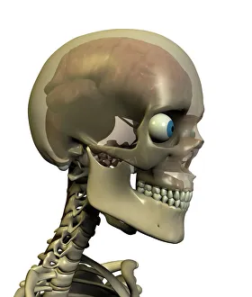
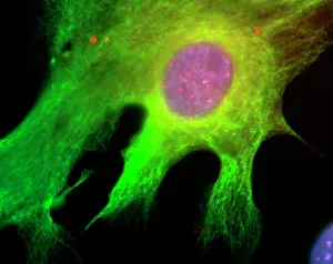
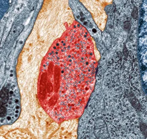
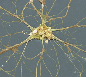
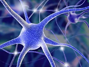
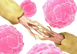
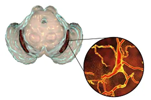
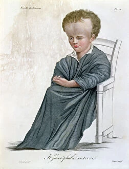
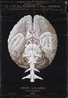
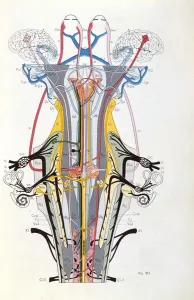
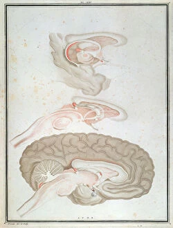
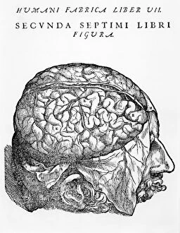
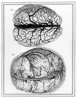
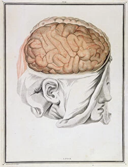
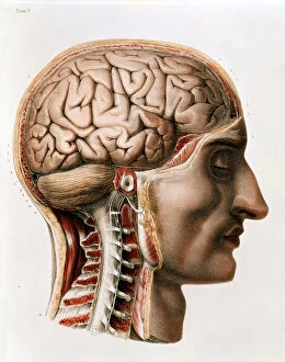
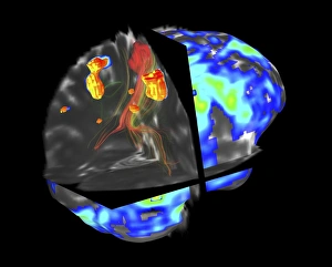
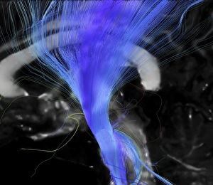
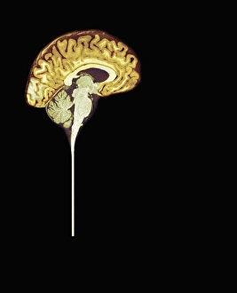
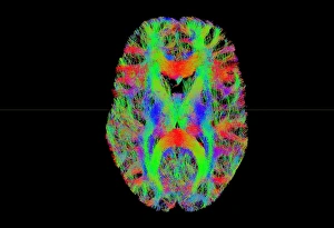
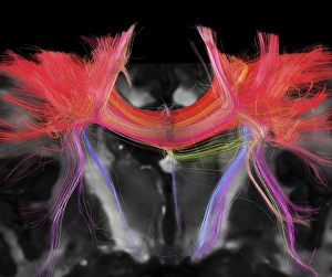
Neurological wonders unravel before our eyes as we delve into the intricate world of the brain. The Motor homunculus model, a representation of the body's motor functions within the brain, unveils the fascinating connection between mind and movement. Cerebellum tissue, captured in a light micrograph, showcases its remarkable structure responsible for coordination and balance. Through Brain fibres revealed by DTI MRI scan C017 / 7099, we witness the complex network that allows information to flow seamlessly throughout our minds. These Brain pathways pave the way for thoughts to travel and actions to be executed with precision. The Brain blood vessels depicted in a stunning 3D angiogram C007 / 1981 remind us of their vital role in nourishing this extraordinary organ. As we explore further, we encounter both Motor and sensory homunculi – visual representations highlighting how different regions of our bodies are represented within specific areas of our brains. Delving deeper into microscopic realms, Nerve and glial cells come alive under a light micrograph. Their intricate structures reveal their crucial roles in transmitting signals and supporting neuronal health. Synapse nerve junctions captured through TEM imagery demonstrate where these incredible cells communicate with one another. Surprisingly, even caffeine crystals make an appearance under a light microscope – reminding us that neurological effects can extend beyond what meets the eye. Zooming closer still reveals Nerve cells observed through SEM imaging; their unique shapes hint at their diverse functions within this awe-inspiring system. Finally, Hippocampus brain tissue takes center stage – known for its involvement in memory formation and spatial navigation. Its intricacies hold secrets waiting to be unlocked by dedicated researchers worldwide. As we uncover these neurological marvels piece by piece, it becomes evident that there is much more than meets the eye when it comes to understanding this enigmatic realm within each one of us - an endless source of fascination awaiting exploration.