Fraud Blocker Skip to main content

Myocardium Collection

The myocardium, also known as the heart muscle, is a vital component of our cardiovascular system

Background imageMyocardium Collection: Heart muscle, confocal light micrograph

Heart muscle, confocal light micrograph
Heart muscle. Confocal light micrograph of a section through cardiac (heart) muscle. Cardiac muscle consists of branching elongated muscle cells

Background imageMyocardium Collection: Anatomy of heart interior, frontal section

Anatomy of heart interior, frontal section

Background imageMyocardium Collection: Gross anatomy of the human heart

Gross anatomy of the human heart

Background imageMyocardium Collection: Heart bypass graft, artwork

Heart bypass graft, artwork
Heart bypass graft. Computer artwork of a heart that has had a blockage of the coronary arteries treated by coronary artery bypass graft (CABG) surgery

Background imageMyocardium Collection: Interior of human heart

Interior of human heart

Background imageMyocardium Collection: Human body showing heart and main circulatory system position

Human body showing heart and main circulatory system position
Transparent human body showing heart and main circulatory system position with internal organs, nervous system, lymphatic system and circulatory system

Background imageMyocardium Collection: Anatomy of human heart, cross section

Anatomy of human heart, cross section with detailed internal structure

Background imageMyocardium Collection: Anterior view of the human heart

Anterior view of the human heart

Background imageMyocardium Collection: Cardiac muscle, TEM

Cardiac muscle, TEM
Cardiac muscle. Coloured transmission electron micrograph (TEM) of cardiac muscle fibrils (orange)from a healthy heart. Mitochondria (pink) supply the muscle cells with energy

Background imageMyocardium Collection: Cardiac muscle and capillary, TEM

Cardiac muscle and capillary, TEM
Cardiac muscle. Coloured transmission electron micrograph (TEM) of cardiac muscle fibrils (purple)from a healthy heart. Mitochondria (green) supply the muscle cells with energy

Background imageMyocardium Collection: Heart with arteries showing cholesterol in one and plaque in the other

Heart with arteries showing cholesterol in one and plaque in the other
Heart with arteries showing cholesterol in one artery and atherosclerotic plaque in the other

Background imageMyocardium Collection: Posterior view of human heart on photo of blood pressure cuff

Posterior view of human heart on photo of blood pressure cuff

Background imageMyocardium Collection: Three dimensional view of human heart, right side

Three dimensional view of human heart, right side

Background imageMyocardium Collection: Insertion of balloon into atherosclerotic artery

Insertion of balloon into atherosclerotic artery

Background imageMyocardium Collection: Arteries on heart showing atherosclerotic plaque in an artery

Arteries on heart showing atherosclerotic plaque in an artery

Background imageMyocardium Collection: 3D rendering of human heart and thoracic veins

3D rendering of human heart and thoracic veins

Background imageMyocardium Collection: Three dimensional view of human heart, back

Three dimensional view of human heart, back

Background imageMyocardium Collection: Heart with pericadium pulled back exposing heart muscle

Heart with pericadium pulled back exposing heart muscle

Background imageMyocardium Collection: Cross section of human heart

Cross section of human heart

Background imageMyocardium Collection: Interior view of heart with detail of muscle cells and atherosclerotic artery

Interior view of heart with detail of muscle cells and atherosclerotic artery. Chest x-ray in background

Background imageMyocardium Collection: Detail of cardiac muscle with blood pressure meter

Detail of cardiac muscle with blood pressure meter

Background imageMyocardium Collection: Human heart covered with pericardium

Human heart covered with pericardium

Background imageMyocardium Collection: Heart valves showing pulmonary valve, mitral valve and tricuspid

Heart valves showing pulmonary valve, mitral valve and tricuspid

Background imageMyocardium Collection: Anterior view of heart and lung on green background

Anterior view of heart and lung on green background

Background imageMyocardium Collection: Comparison of normal heart versus heart with a patent foramen ovale

Comparison of normal heart versus heart with a patent foramen ovale

Background imageMyocardium Collection: Conceptual image of human heart

Conceptual image of human heart

Background imageMyocardium Collection: Internal view of the human heart

Internal view of the human heart

Background imageMyocardium Collection: Interior of human heart showing atria and ventricles

Interior of human heart showing atria and ventricles

Background imageMyocardium Collection: Enlarged left ventricle of the human heart

Enlarged left ventricle of the human heart

Background imageMyocardium Collection: Three dimensional view of human heart, left side

Three dimensional view of human heart, left side

Background imageMyocardium Collection: Illustration of human heart

Illustration of human heart

Background imageMyocardium Collection: Heart muscle, fluorescence micrograph C016 / 8483

Heart muscle, fluorescence micrograph C016 / 8483
Heart muscle. Fluorescence deconvolution micrograph of a section through heart tissue, showing the angular distribution of the myocardium (cardiac muscle) fibres (green)

Background imageMyocardium Collection: Heart muscle, fluorescence micrograph C016 / 8481

Heart muscle, fluorescence micrograph C016 / 8481
Heart muscle. Fluorescence deconvolution micrograph of a section through heart tissue, showing blood vessels (oval, centre-left and upper centre) running through the myocardium (cardiac muscle)

Background imageMyocardium Collection: Heart muscle, fluorescence micrograph C016 / 8478

Heart muscle, fluorescence micrograph C016 / 8478
Heart muscle. Fluorescence deconvolution micrograph of a section through heart tissue, showing a blood vessel (diagonal, centre) running through the myocardium (cardiac muscle, green)

Background imageMyocardium Collection: Heart muscle, fluorescence micrograph C016 / 8477

Heart muscle, fluorescence micrograph C016 / 8477
Heart muscle. Fluorescence deconvolution micrograph of a section through heart tissue, showing a blood vessel (blue, diagonal, centre) running through the myocardium (cardiac muscle, green)

Background imageMyocardium Collection: Pericardium, TEM

Pericardium, TEM
Pericardium. Coloured transmission electron micrograph (TEM) of a section through the serous pericardium. The pericardium is a double-walled sac surrounding the heart

Background imageMyocardium Collection: Electrical conduction in the heart, artwork

Electrical conduction in the heart, artwork. Each heartbeat is initiated by an electrical impulse from the sinoatrial (SA) node (green oval) in the right atrium (upper heart chamber)

Background imageMyocardium Collection: Heart bypass grafts

Heart bypass grafts. Artwork of a heart that has had a blockage of the coronary arteries treated by coronary artery bypass graft (CABG) surgery

Background imageMyocardium Collection: Heart blood vessel wall, TEM

Heart blood vessel wall, TEM

Background imageMyocardium Collection: Heart muscle, SEM

Heart muscle, SEM
Heart muscle. Coloured scanning electron micrograph (SEM) of pectinate muscles in the wall of the right atrium of the heart

Background imageMyocardium Collection: Damaged cardiac tissue, artwork

Damaged cardiac tissue, artwork
Damaged cardiac tissue. Artwork of damaged heart fibres and cells in heart muscle tissue (myocardium). This can occur due to reduced blood supply (ischaemia)

Background imageMyocardium Collection: Cardiac muscle, artwork

Cardiac muscle, artwork
Cardiac muscle. Computer artwork of cardiac muscle fibres. Cardiac muscle is under subconscious control and continuously contracts to pump blood around the body without tiring

Background imageMyocardium Collection: Trabeculae carneae in the heart, SEM

Trabeculae carneae in the heart, SEM
Trabeculae carneae in the left ventricle of the heart, coloured scanning electron micrograph (SEM). Endocardial connective tissue is also seen

Background imageMyocardium Collection: Heart valve and strings, SEM

Heart valve and strings, SEM
Heart valve and strings, coloured scanning electron micrograph (SEM). This is the mitral valve (upper right), controlling the blood flow between two of the hearts chambers

Background imageMyocardium Collection: Heart valves and strings, SEM

Heart valves and strings, SEM
Heart valves and strings, coloured scanning electron micrograph (SEM). This dissection shows both the mitral valve (centre) and the aortic valve (upper centre)


All Professionally Made to Order for Quick Shipping

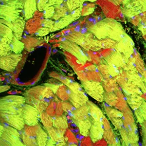
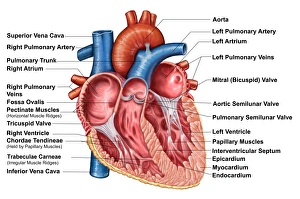
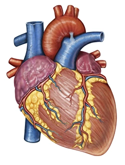
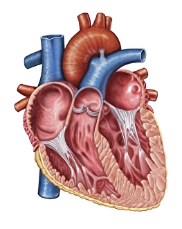
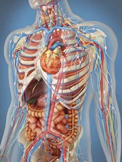
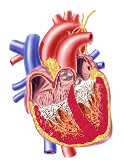
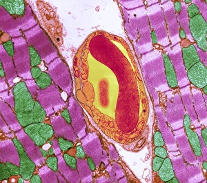
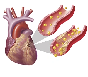
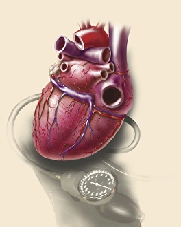
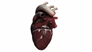
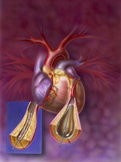
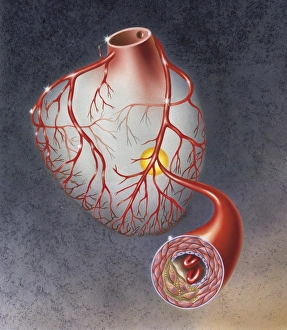
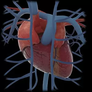
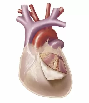
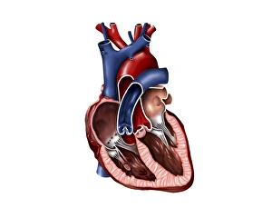
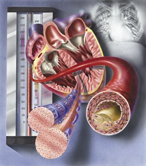
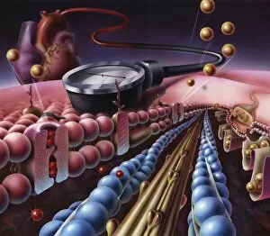
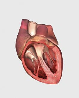
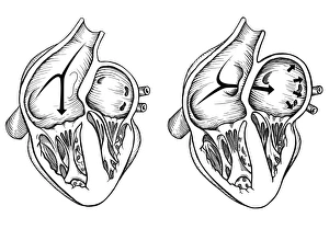
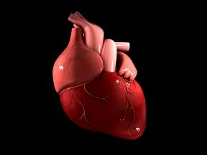
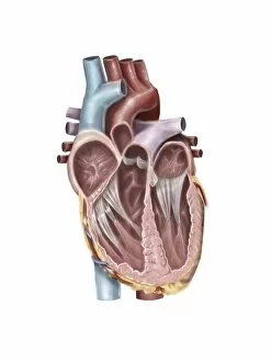
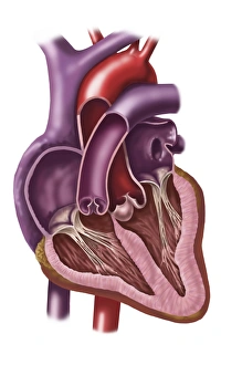
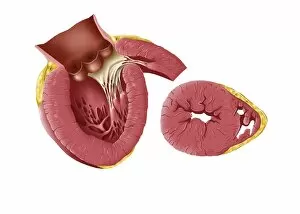
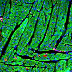
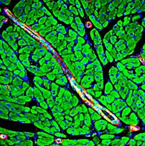
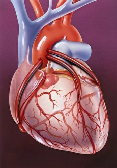
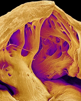
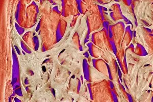
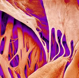
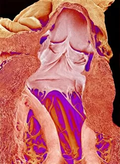
The myocardium, also known as the heart muscle, is a vital component of our cardiovascular system. This confocal light micrograph showcases its intricate structure and highlights its importance in maintaining a healthy heart. When we explore the anatomy of the heart interior through a frontal section, we can truly appreciate the complexity and beauty of this organ. The myocardium stands out prominently, surrounded by other essential structures that ensure proper functioning. In gross anatomy images of the human heart, we witness how the myocardium forms a significant part of this remarkable organ. Its strength and resilience enable it to pump blood efficiently throughout our bodies. Artwork depicting a heart bypass graft reminds us of the incredible medical advancements that help restore blood flow when necessary. The myocardium plays an integral role in these procedures, ensuring oxygen-rich blood reaches every corner of our body. Peering into the interior of a human heart reveals not only its chambers but also emphasizes how crucially positioned it is within our main circulatory system. The myocardium's contractions propel life-sustaining blood to all parts of our body without fail. Examining cardiac muscle at a microscopic level using transmission electron microscopy (TEM) allows us to marvel at its unique cellular structure. These specialized cells work tirelessly together to maintain optimal cardiac function day after day. The close relationship between cardiac muscle and capillaries becomes evident under TEM imaging. Capillaries intricately weave around individual cardiac muscle fibers, providing them with nutrients and oxygen while removing waste products - an exquisite symbiotic partnership within our hearts. A three-dimensional view from the front provides us with yet another perspective on this magnificent organ. It showcases how each component fits seamlessly together - including the indispensable myocardium - enabling efficient circulation throughout our bodies. Visualizing arteries showing cholesterol buildup in one image and plaque formation in another serves as an important reminder about cardiovascular health challenges faced by many individuals today. Understanding how these conditions impact both overall heart health and specifically the myocardium is crucial for prevention and treatment.