
Murine Collection
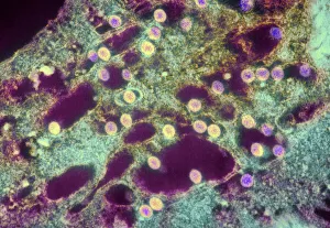
"Exploring the World of Murine: From Rift Valley Fever Virus to Neural Stem Cells" Delving into the microscopic realm, we witness the Rift Valley fever virus under TEM


All Professionally Made to Order for Quick Shipping
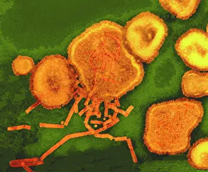
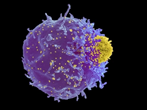
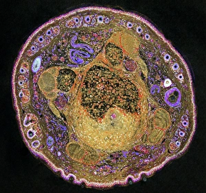
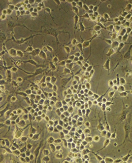
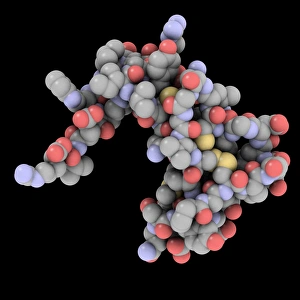
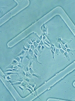
"Exploring the World of Murine: From Rift Valley Fever Virus to Neural Stem Cells" Delving into the microscopic realm, we witness the Rift Valley fever virus under TEM, revealing its intricate structure and potential implications. Paramyxovirus particles captured under TEM showcase their unique morphology, shedding light on their role in various diseases. The captivating Murine Minute Virus capsid takes center stage as it unveils its elegant architecture through advanced imaging techniques. An illustrated depiction of a mouse (C018/0739) introduces us to these fascinating creatures that have played a crucial role in scientific research for decades. Peering into a prepared specimen (C018/0318) of a mouse embryo provides an awe-inspiring glimpse into the early stages of life and development. Returning to the mesmerizing Murine Minute Virus capsid, we marvel at its complexity and significance within viral biology. Molecular models unveil the intricacies of Mengovirus capsids, offering insights into their structural composition and potential therapeutic targets. A striking SEM image showcases Mouse Leukemia Virus interacting with T-cells (C017/8308), unraveling critical aspects of viral-host interactions in disease progression. Through a light micrograph, we explore the detailed anatomy of a mouse tail – an essential feature contributing to locomotion and sensory perception. Zooming in on hair follicles using SEM reveals their remarkable structure and function within mammalian skin – nature's exquisite design for insulation and protection. Shifting our focus towards neural stem cells opens up new possibilities for understanding brain development, regeneration, and neurodegenerative disorders like never before. In this captivating journey through murine wonders - from viruses to embryonic development.