
Mouthparts Collection
"Mouthparts: Unveiling the Intricate World of Nature's Feeding Mechanisms" Discovering the mesmerizing world of mouthparts


All Professionally Made to Order for Quick Shipping
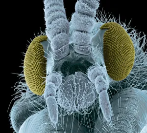
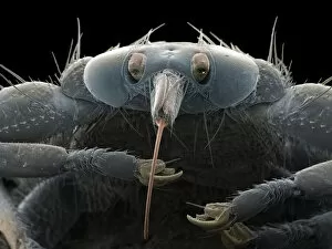
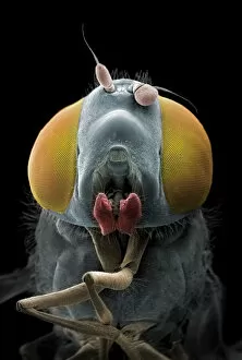
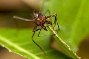
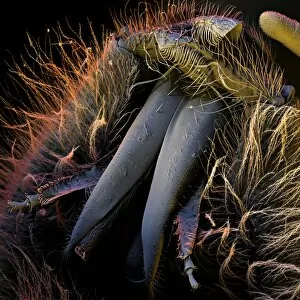
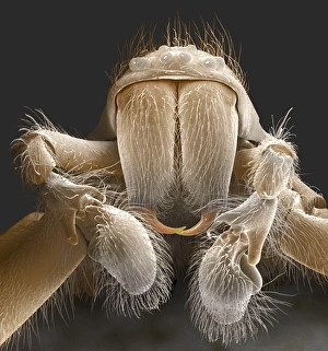
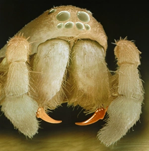
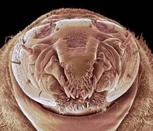
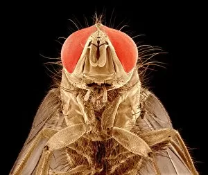
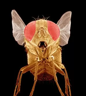
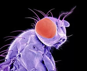
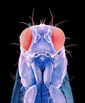
"Mouthparts: Unveiling the Intricate World of Nature's Feeding Mechanisms" Discovering the mesmerizing world of mouthparts, as captured through a colored scanning electron micrograph of a fly head. These tiny structures hold secrets that both fascinate and deceive. Intriguingly irresistible, the liar within these mouthparts is unveiled. A leaf-cutter ant from Pacaya-Samiria NR in Peru showcases its impeccable cutting skills, highlighting the precision with which it wields its jaw-like mandibles. Delving deeper into this microscopic realm, we encounter a maggot head under SEM C014/1449. Its intricate anatomy leaves us awestruck, reminding us that beauty can be found even in unexpected places. Artistry takes form as we explore spider anatomy depicted through an exquisite artwork. The complexity of their mouthparts serves as a testament to nature's ingenuity and adaptability. Moving on to honey bees' mouths under SEM C016/8004, we witness their delicate yet efficient design for sipping nectar and collecting pollen. Their vital role in pollination becomes evident as we marvel at their specialized proboscis. The bullet ant (C018/2481) and leafcutter ant (C018/2390) make appearances next, showcasing diverse adaptations for survival within their respective ecosystems. Their formidable jaws remind us of nature's endless variety and resourcefulness. Picture No. 11675186 brings forth the head of an Anopheles mosquito, revealing female mouthparts responsible for transmitting diseases through biting humans. This reminder emphasizes the importance of understanding these mechanisms to combat vector-borne illnesses effectively. From flies to ants, maggots to spiders – this captivating journey through various species' mouthparts sheds light on their remarkable diversity and functionality within our natural world.