
Mouthpart Collection
"The Liar: Unveiling the Irresistible World of Mouthparts" Delve into the intriguing world of mouthparts, where truth and deception intertwine


All Professionally Made to Order for Quick Shipping
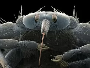
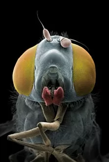
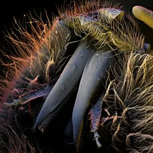
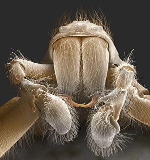
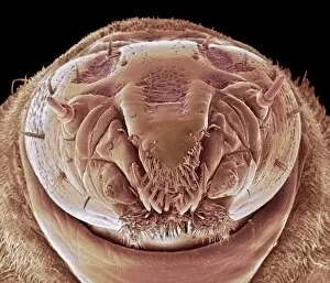
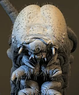
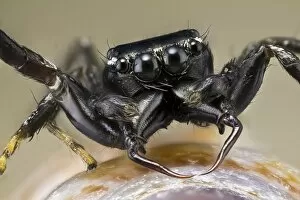
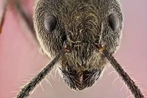
"The Liar: Unveiling the Irresistible World of Mouthparts" Delve into the intriguing world of mouthparts, where truth and deception intertwine. From the deceptive liar to the irresistible allure, these tiny structures hold captivating secrets waiting to be unraveled. Witness the grotesque beauty of Maggot Head under SEM C014 / 1449, as it showcases its unique mouthpart structure designed for survival. Marvel at Spider Anatomy Artwork that intricately portrays their diverse feeding mechanisms. Explore Honey Bee Mouth under SEM C016 / 8004, a masterpiece of nature's engineering marvels. Discover how this intricate apparatus enables them to collect nectar and pollen with utmost precision. Beware the Bullet Ant C018 / 2481 - its formidable a weapon in disguise. Witness Leafcutter Ant C018 / 2390's efficient mandibles as they slice through foliage like miniature scissors. Uncover Spider Mouthparts under SEM Z430 / 0436; each species adapted for different prey capture strategies. Observe House Spider's intricate mouthparts under SEM, revealing their hidden hunting prowess. Marvel at Fruit Fly Proboscis under SEM; an elegant tool used for sipping on sweet nectar or puncturing fruits with surgical precision. Encounter Wandering Spider C014 / 9804 - its venomous fangs ready to strike fear into any trespasser. Lastly, meet Harvestman – an enigmatic creature whose delicate-looking mouthpart belies its predatory nature. These fascinating beings challenge our understanding of evolution and adaptation within arachnid communities. Join us on this journey through magnified wonders where mouths become tools for survival, deceitful tactics emerge from unlikely sources, and beauty lies in even the most unexpected places. Prepare to be captivated by these remarkable creatures' extraordinary tales told through their mesmerizing mouthparts.