Fraud Blocker Skip to main content

Molecular Biology Collection (page 13)

"Molecular Biology: Unveiling the Secrets of Life at a Microscopic Level" Amitriptyline Antidepressant Molecule: Exploring the Chemical Pathways to Mental Well-being

Background imageMolecular Biology Collection: Infectious bursal disease virus capsid

Infectious bursal disease virus capsid, molecular model. This avian virus infects the bursa of Fabricius (specialised bird immune organ) in young chickens, and can cause high mortality rates

Background imageMolecular Biology Collection: Bacteriorhodopsin protein

Bacteriorhodopsin protein. Molecular model showing the structure of bacteriorhodopsin (bR), a protein found in primitive micro-organisms known as Archaea. This protein acts as a proton pump

Background imageMolecular Biology Collection: Bacteriophage RNA, molecular model

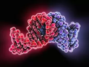
Bacteriophage RNA, molecular model
Bacteriophage RNA. Molecular model showing the structure of a loop of the genetic material RNA (ribonucleic acid) from a bacteriophage. Bacteriophages are viruses that infect bacteria

Background imageMolecular Biology Collection: Enterovirus particle C014 / 4900

Enterovirus particle C014 / 4900
Enterovirus particle. Computer artwork of an enterovirus particle (virion), showing the structure of the capsid (outer shell)

Background imageMolecular Biology Collection: Bacteriophage connector protein

Bacteriophage connector protein. Molecular model showing the structure of a bacteriophage head-tail connector protein. Bacteriophages are viruses that infect bacteria

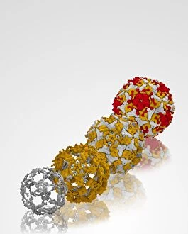
Background imageMolecular Biology Collection: Enterovirus capsid proteins structure C014 / 4897

Enterovirus capsid proteins structure C014 / 4897
Enterovirus capsid proteins structure. Computer artwork showing how the four component proteins (VP1 to VP4) of an enterovirus particle (virion) interlock to form the capsid (outer shell)

Background imageMolecular Biology Collection: Cholecystokinin-8 molecule C014 / 4895

Cholecystokinin-8 molecule C014 / 4895
Cholecystokinin-8 molecule. Computer model showing the structure of the terminal fragment of a molecule of the hormone cholecystokinin-8 (CCK-8)
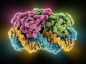
Background imageMolecular Biology Collection: Enterovirus capsid proteins structure C014 / 4896

Enterovirus capsid proteins structure C014 / 4896
Enterovirus capsid proteins structure. Computer artwork showing how the four component proteins (VP1 to VP4) of an enterovirus particle (virion) interlock to form the capsid (outer shell)

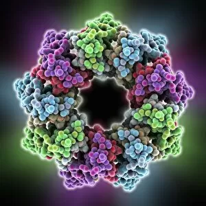
Background imageMolecular Biology Collection: MscS ion channel protein structure

MscS ion channel protein structure. Molecular model showing the protein structure of a Mechanosensitive Channel of Small Conductance (MscS) from an Escherichia coli bacterium

Background imageMolecular Biology Collection: Bacteriophage capsid protein shell

Bacteriophage capsid protein shell. Molecular model showing the partial shell structure of a bacteriophage capsid based on one of its coat proteins

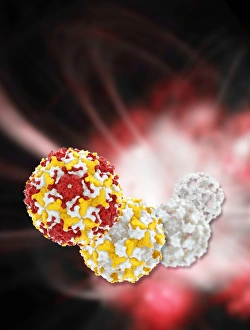
Background imageMolecular Biology Collection: Enterovirus particle C014 / 4898

Enterovirus particle C014 / 4898
Enterovirus particle. Computer artwork of an enterovirus particle (virion), showing the structure of the capsid (outer shell)

Background imageMolecular Biology Collection: Bacterial outer membrane protein molecule C014 / 4949

Bacterial outer membrane protein molecule C014 / 4949
Bacterial outer membrane protein molecule. Computer model showing the secondary structure of a molecule of outer membrane transporter FecA protein from Escherichia coli (E. coli) bacteria
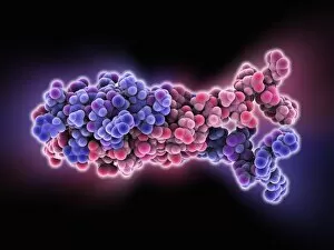
Background imageMolecular Biology Collection: Cholecystokinin-8 molecule C014 / 4894

Cholecystokinin-8 molecule C014 / 4894
Cholecystokinin-8 molecule. Computer model showing the structure of the terminal fragment of a molecule of the hormone cholecystokinin-8 (CCK-8)

Background imageMolecular Biology Collection: Enterovirus particles C014 / 4899

Enterovirus particles C014 / 4899
Enterovirus particles. Computer artwork of enterovirus particles (virion). Enteroviruses are a genus of non-enveloped positive-sense single-stranded RNA viruses associated with several human

Background imageMolecular Biology Collection: Influenza proton pump, molecular model

Influenza proton pump, molecular model
Influenza proton pump. Molecular model showing the protein structure of a proton pump from an influenza virus. Proton pumps are membrane proteins that move protons across a cell membrane

Background imageMolecular Biology Collection: DNA quadruplex, molecular model

DNA quadruplex, molecular model. This dimeric quadruplex of DNA (deoxyribonucleic acid) is thought to form as part of telomeres

Background imageMolecular Biology Collection: Bone morphogenetic protein complex

Bone morphogenetic protein complex, molecular model. Bone Morphogenetic Protein-7 (BMP-7, blue) in complex with the secreted antagonist Noggin (pink)

Background imageMolecular Biology Collection: Light-harvesting protein complex

Light-harvesting protein complex, molecular model. Peripheral light-harvesting protein complex from the purple bacterium Rhodopseudomonas acidophila

Background imageMolecular Biology Collection: Biotin ligase enzyme, molecular model

Biotin ligase enzyme, molecular model. This enzyme is a protein formed from 268 amino acids and two chains (green and pink)

Background imageMolecular Biology Collection: B-Z junction in DNA, molecular model

B-Z junction in DNA, molecular model. Deoxyribonucleic acid (DNA) occurs in three forms, A-DNA, B-DNA and Z-DNA. The first two are right-handed, with B-DNA being the more common form

Background imageMolecular Biology Collection: Cat allergen protein, molecular model C015 / 3962

Cat allergen protein, molecular model C015 / 3962
Cat allergen protein. Molecular model of the tetrameric form of the major cat allergen fel d 1 (Felis domesticus allergen 1)

Background imageMolecular Biology Collection: Bacterial RNA plasmid loop-loop complex

Bacterial RNA plasmid loop-loop complex, molecular model. This strand of ribonucleic acid (RNA) is part of a plasmid, the loop of genetic material found in bacterial cells

Background imageMolecular Biology Collection: Cat allergen protein, molecular model

Cat allergen protein, molecular model
Cat allergen protein. Molecular model of the tetrameric form of the major cat allergen fel d 1 (Felis domesticus allergen 1)

Background imageMolecular Biology Collection: Methane monooxygenase enzyme

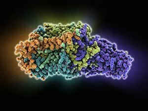
Methane monooxygenase enzyme, molecular model. This is the particulate methane monooxygenase (pMMO) form of this metalloenzyme, an integral membrane protein that contains copper and zinc

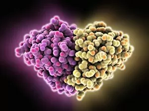
Background imageMolecular Biology Collection: FP2 malaria protease enzyme complex

FP2 malaria protease enzyme complex, molecular model. This complex consists of the falcipain-2 (FP2) protease enzyme (purple, right) bound to a cystatin (orange, left), a form of protease inhibitor

Background imageMolecular Biology Collection: Follicle-stimulating hormone complex C015 / 0945

Follicle-stimulating hormone complex C015 / 0945
Follicle-stimulating hormone (FSH) complex with receptor, molecular model. FSH helps to regulate human sexual development and reproductive processes. In females, it acts on follicles in the ovaries

Background imageMolecular Biology Collection: Follicle-stimulating hormone complex C015 / 0944

Follicle-stimulating hormone complex C015 / 0944
Follicle-stimulating hormone (FSH) complex with receptor, molecular model. FSH helps to regulate human sexual development and reproductive processes. In females, it acts on follicles in the ovaries

Background imageMolecular Biology Collection: Glutamate transporter protein

Glutamate transporter protein, molecular model. This is a membrane protein that facilitates the uptake of glutamate by a cell, thus playing an important role in neurology in higher organisms

Background imageMolecular Biology Collection: Purple bacterium photosynthesis centre

Purple bacterium photosynthesis centre, molecular model. Purple bacteria are phototrophic bacteria that produce energy through photosynthesis

Background imageMolecular Biology Collection: Oestrogen related receptor-DNA complex

Oestrogen related receptor-DNA complex. Molecular model of human estrogen related receptor-2 (heRR-2, purple) binding to a strand of DNA (deoxyribonucleic acid, red and yellow-green)

Background imageMolecular Biology Collection: Lambda repressor-operator complex

Lambda repressor-operator complex. Molecular model of the lambda repressor protein (red and green) binding to a region of DNA (deoxyribonucleic acid, orange and blue) known as the lambda operator

Background imageMolecular Biology Collection: H-NS chromatin-structuring protein

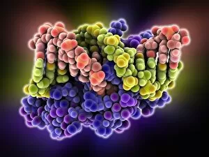
H-NS chromatin-structuring protein. Molecular model of the oligomerization domain of the H-NS protein from the Escherichia coli bacterium. This dimeric molecule folds in on itself, as shown here

Background imageMolecular Biology Collection: Chloride ion channel, molecular model

Chloride ion channel, molecular model. This is a ClC ion channel. Its role is to mediate the flow of chloride ions across cell membranes

Background imageMolecular Biology Collection: Repair protein and DNA, molecular model

Repair protein and DNA, molecular model
Repair protein and DNA. Molecular model of the Ku heterodimer (grey, blue and purple) bound to a strand of DNA (deoxyribonucleic acid, orange and green) as part of the repair process

Background imageMolecular Biology Collection: Max transcription factor-DNA complex

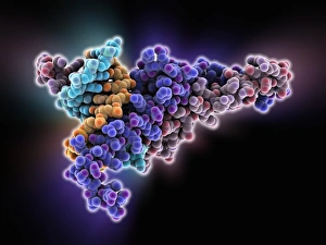
Max transcription factor-DNA complex. Molecular model of the Max transcription factor (purple and red) bound to a strand of DNA (deoxyribonucleic acid, light blue and orange)

Background imageMolecular Biology Collection: Antibody fragment-lysozyme complex

Antibody fragment-lysozyme complex
fab d1.3, , chicken egg white lysozyme, protein, biomolecule, macromolecule, lysozyme, enzyme, fab, antigen, antibody, biochemistry, biology, molecular biology, proteomics, artwork, illustration

Background imageMolecular Biology Collection: Bacterial regulator-DNA complex

Bacterial regulator-DNA complex. Molecular model of a complex formed between a bacterial regulator called SarA (orange and brown) and a fragment of DNA (pink and yellow-green strands)

Background imageMolecular Biology Collection: Bacterial protein-chaperone complex

Bacterial protein-chaperone complex. Molecular model of a bacterial effector protein binding to a chaperone protein that helps prevent keep the bacterial protein in an unfolded or partially folded

Background imageMolecular Biology Collection: Hin recombinase-DNA complex

Hin recombinase-DNA complex. Molecular model of the Hin recombinase protein (pink) bound to a double helix (green and orange) strand of DNA (deoxyribonucleic acid)

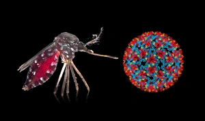
Background imageMolecular Biology Collection: Aedes mosquito and Chikungunya virus

Aedes mosquito and Chikungunya virus. Asian tiger mosquito (Aedes albopictus) mosquito next to a Chikungunya virus particle (virion). The Chikungunya virus is transmitted by Aedes sp


All Professionally Made to Order for Quick Shipping

"Molecular Biology: Unveiling the Secrets of Life at a Microscopic Level" Amitriptyline Antidepressant Molecule: Exploring the Chemical Pathways to Mental Well-being. Watson and Crick, DNA Discovers: Pioneers Who Unraveled the Double Helix Structure, Revolutionizing Genetics. Creatine Amino Acid Molecule: Fueling Muscles and Unlocking Athletic Performance Potential. Microscopic View of Human Respiratory Syncytial Virus: Understanding the Culprit Behind Common Respiratory Infections. Embryo Development 24-36 Hours After Fertilization: Witnessing the Miracle of Life from Conception to Early Growth. Zinc Fingers Bound to a DNA Strand: Decoding Genetic Instructions for Cellular Functions and Disease Prevention. SARS Coronavirus Protein: Investigating Viral Mechanisms for Effective Control and Treatment Strategies. Vioxx Drug Molecule: Analyzing Medications' Molecular Structures for Enhanced Safety and Efficacy in Pain Management. Levofloxacin Antibiotic Molecule: Combating Bacterial Infections with Precision-targeted Therapies. Electrophoresis of RNA: Separating Genetic Material to Study Gene Expression Patterns in Health and Disease Mitochondrial DNA - The Powerhouses Within Our Cells Holding Clues to Evolutionary History Cone Snail Venom Component Molecule.