
Mineralised Collection
"Unveiling the Marvels of Mineralised Microcosms: Exploring Calcareous Phytoplankton and Diatoms through SEM" Step into a world unseen by the naked eye


All Professionally Made to Order for Quick Shipping
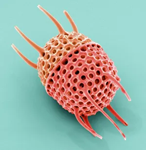
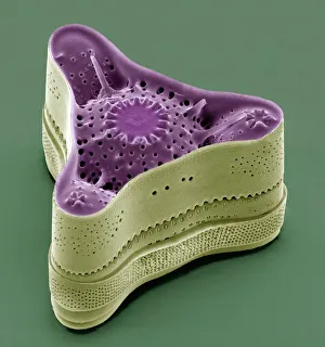
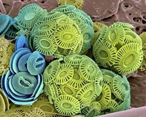
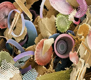
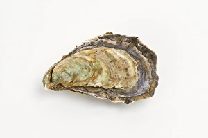
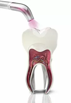
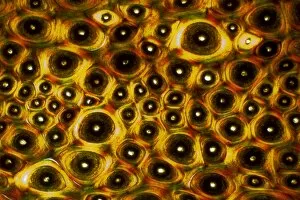
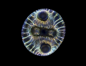
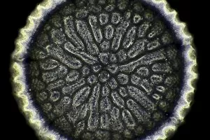
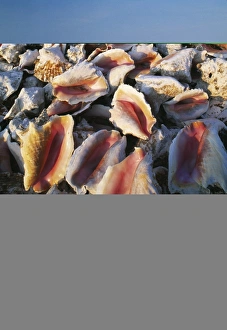
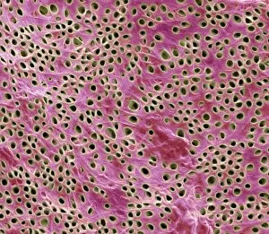
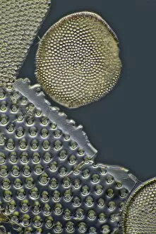
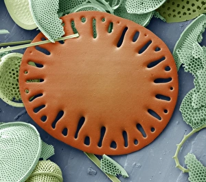
"Unveiling the Marvels of Mineralised Microcosms: Exploring Calcareous Phytoplankton and Diatoms through SEM" Step into a world unseen by the naked eye, where intricate structures and fascinating formations await. In this captivating journey, we delve into the realm wonders, starting with calcareous phytoplankton. Under the scanning electron microscope (SEM), these tiny organisms reveal their ornate shells, showcasing nature's artistry at its finest. Moving forward, our focus shifts to diatoms - another group of mesmerizing microorganisms. Their delicate frustules come alive under SEM's magnifying lens, unveiling intricate patterns that resemble miniature works of art. Each diatom species boasts its own unique design, leaving us in awe of their diversity and complexity. As we continue our exploration, fossilized diatoms take center stage. Preserved over time in sedimentary rocks, these ancient remnants offer glimpses into Earth's past ecosystems. Through SEM imaging techniques, we unravel their secrets and gain insights into prehistoric environments long gone. But minerals don't limit themselves to microscopic life forms alone; they also leave an indelible mark on petrified wood. Witnessing the transformation from organic matter to stone is truly remarkable as tree rings become infused with minerals over centuries or even millennia. Returning to diatoms once more – each new image captured by SEM reveals astonishing details previously unseen. The sheer variety in shape and structure continues to astound us as we uncover more hidden treasures within this diverse group of microalgae. Beyond natural phenomena lies unexpected beauty found in unlikely places - like Pacific oysters adorned with shimmering mineral deposits that enhance their already exquisite appearance. Lastly, stepping away from biological marvels for a moment brings us to dental filling polymerization artwork C014 / 2025 – a fusion of science and creativity showcased under SEM's watchful eye. This artistic representation captures the intricate process of dental filling polymerization, highlighting the intersection between science and art.