Fraud Blocker Skip to main content

Microorganisms Collection (page 8)

Microorganisms, the tiny wonders of life that exist all around us, are both fascinating and formidable

Background imageMicroorganisms Collection: Netrium desmids, light micrograph

Netrium desmids, light micrograph
Netrium desmids. Polarised light micrograph of Netrium sp. desmid green alga. Desmids are a common group of freshwater single-celled algae that have intricate cell walls

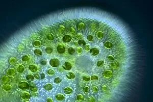
Background imageMicroorganisms Collection: Vorticella protozoan, light micrograph

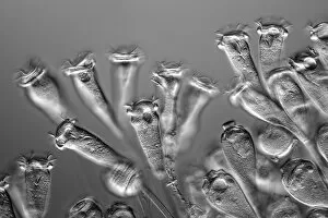
Vorticella protozoan, light micrograph
Vorticella protozoan. Rheinberg illuminated light micrograph of a Vorticella sp. ciliate protozoan. This single-celled organism consists of a bell-shaped head

Background imageMicroorganisms Collection: Cosmarium desmids, light micrograph

Cosmarium desmids, light micrograph
Phase contrast, magnification: 200 X on APS-C sensor

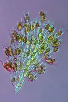
Background imageMicroorganisms Collection: Golden algae, light micrograph

Golden algae, light micrograph
Golden algae. Polarised light micrograph of a colony of Dinobryon sp. golden algae (chrysophytes). Magnification: x200 when printed 10 centimetres wide

Background imageMicroorganisms Collection: Paramecium bursaria protozoan, light micr

Paramecium bursaria protozoan, light micr
Paramecium bursaria protozoan, differential interference contrast light micrograph. This ciliate protozoan inhabits freshwater, where it feeds mainly on bacteria

Background imageMicroorganisms Collection: Euastrum desmids, light micrograph

Euastrum desmids, light micrograph
Euastrum desmids. Polarised light micrograph of Euastrum sp. desmid green algae. Desmids are a common group of freshwater single-celled algae that have intricate cell walls

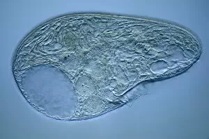
Background imageMicroorganisms Collection: Blepharisma protozoan, light micrograph

Blepharisma protozoan, light micrograph
Blepharisma protozoan. Differential interference contrast micrograph of a Blepharisma japonicum protozoan. These ciliate protozoa move by beating their cilia

Background imageMicroorganisms Collection: Blepharisma protozoa, light micrograph

Blepharisma protozoa, light micrograph
Blepharisma protozoa. Differential interference contrast micrograph of Blepharisma japonicum protozoa. These ciliate protozoa move by beating their cilia

Background imageMicroorganisms Collection: Micrasterias desmids, light micrograph

Micrasterias desmids, light micrograph
Micrasterias desmids. Polarised light micrograph of Micrasterias truncata desmid green algae. Desmids are a common group of freshwater single-celled algae that have intricate cell walls

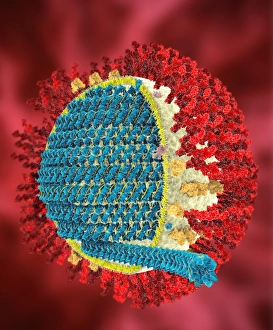
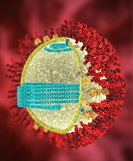
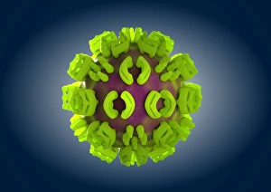
Background imageMicroorganisms Collection: MERS coronavirus, artwork C016 / 3611

MERS coronavirus, artwork C016 / 3611
MERS coronavirus proteins. Cutaway computer model showing the protein structure of a MERS coronavirus particle (virion). MERS (Middle East respiratory syndrome)

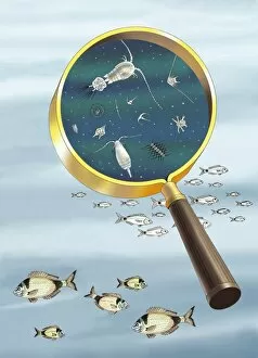
Background imageMicroorganisms Collection: Plankton and fish, artwork C016 / 3448

Plankton and fish, artwork C016 / 3448
Plankton and fish. Artwork of plankton as viewed under a magnifying glass, with seabream (Diplodus vulgaris) fish

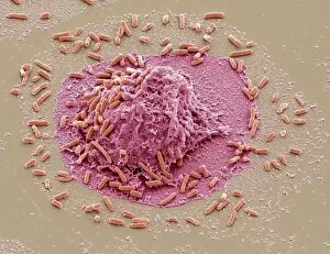
Background imageMicroorganisms Collection: E. coli induced cell death, SEM C016 / 3078

E. coli induced cell death, SEM C016 / 3078
E. coli induced cell death. Coloured scanning electron micrograph (SEM) of a macrophage white blood cell (centre) that is being destroyed by toxins released by Escherichia coli (E)

Background imageMicroorganisms Collection: E. coli induced cell death, SEM C016 / 3077

E. coli induced cell death, SEM C016 / 3077
E. coli induced cell death. Coloured scanning electron micrograph (SEM) of a macrophage white blood cell (centre) that is being destroyed by toxins released by Escherichia coli (E)

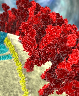
Background imageMicroorganisms Collection: SARS coronavirus proteins, artwork C016 / 3056

SARS coronavirus proteins, artwork C016 / 3056
SARS coronavirus proteins. Computer model showing the spike proteins (red) of a SARS coronavirus particle (virion). SARS (severe acute respiratory syndrome)

Background imageMicroorganisms Collection: SARS coronavirus, artwork C016 / 3055

SARS coronavirus, artwork C016 / 3055
SARS coronavirus proteins. Computer artwork of SARS coronavirus particles (virions). SARS (severe acute respiratory syndrome)

Background imageMicroorganisms Collection: SARS coronavirus, artwork C016 / 3054

SARS coronavirus, artwork C016 / 3054
SARS coronavirus proteins. Cutaway computer model showing the protein structure of a SARS coronavirus particle (virion). SARS (severe acute respiratory syndrome)

Background imageMicroorganisms Collection: SARS coronavirus, artwork C016 / 3053

SARS coronavirus, artwork C016 / 3053
SARS coronavirus proteins. Cutaway computer model showing the protein structure of a SARS coronavirus particle (virion). SARS (severe acute respiratory syndrome)

Background imageMicroorganisms Collection: SARS coronavirus proteins, artwork C016 / 3052

SARS coronavirus proteins, artwork C016 / 3052
SARS coronavirus proteins. Computer model showing the proteins of a SARS coronavirus particle (virion). SARS (severe acute respiratory syndrome)

Background imageMicroorganisms Collection: Vorticella protozoa, light micrograph C016 / 3042

Vorticella protozoa, light micrograph C016 / 3042
Vorticella protozoa. Differential interference contrast (DIC) light micrograph of Vorticella sp. ciliate protozoa. These single-celled organisms consist of a bell-shaped head

Background imageMicroorganisms Collection: Vorticella protozoan, light micrograph C016 / 3043

Vorticella protozoan, light micrograph C016 / 3043
Vorticella protozoan. Differential interference contrast (DIC) light micrograph of a Vorticella sp. ciliate protozoan. This single-celled organism consists of a bell-shaped head

Background imageMicroorganisms Collection: Vorticella protozoa, light micrograph C016 / 3041

Vorticella protozoa, light micrograph C016 / 3041
Vorticella protozoa. Differential interference contrast (DIC) light micrograph of Vorticella sp. ciliate protozoa. These single-celled organisms consist of a bell-shaped head

Background imageMicroorganisms Collection: Vorticella protozoa, light micrograph C016 / 3040

Vorticella protozoa, light micrograph C016 / 3040
Vorticella protozoa. Differential interference contrast (DIC) light micrograph of Vorticella sp. ciliate protozoa. These single-celled organisms consist of a bell-shaped head

Background imageMicroorganisms Collection: Satellite tobacco necrosis virus C014 / 0005

Satellite tobacco necrosis virus C014 / 0005
Satellite tobacco necrosis virus. Computer artwork showing the surface structure of a tobacco necrosis satellivirus particle (virion)

Background imageMicroorganisms Collection: Canine parvovirus particle C013 / 9966

Canine parvovirus particle C013 / 9966
Canine parvovirus particle. Computer artwork showing the structure of the outer protein coat (capsid) of a canine parvovirus type 2 particle (virion)

Background imageMicroorganisms Collection: Salmonella bacteria, artwork C013 / 7809

Salmonella bacteria, artwork C013 / 7809
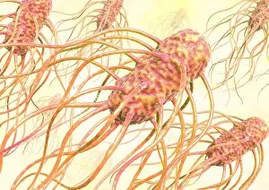
Salmonella bacteria, computer artwork. Salmonella sp. bacteria are gram-negative rod-shaped bacteria that have flagella (hair-like structures) that they use for locomotion

Background imageMicroorganisms Collection: Salmonella bacteria, artwork C013 / 7808

Salmonella bacteria, artwork C013 / 7808
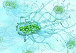
Salmonella bacteria, computer artwork. Salmonella sp. bacteria are gram-negative rod-shaped bacteria that have flagella (hair-like structures) that they use for locomotion

Background imageMicroorganisms Collection: Bird flu virus particle, artwork C013 / 4650

Bird flu virus particle, artwork C013 / 4650
Bird flu virus particle. Computer artwork showing the internal and external structures of an influenza (flu) A subtype H5N1 (bird flu) virus particle (virion)

Background imageMicroorganisms Collection: Ross River virus particle, artwork C013 / 4641

Ross River virus particle, artwork C013 / 4641
Ross River virus particle. Computer artwork showing the outer structure of a Ross River virus (RRV) particle (virion). RRV is a small encapsulated single-strand RNA alphavirus endemic to Australia

Background imageMicroorganisms Collection: Marburg virus, artwork C013 / 4633

Marburg virus, artwork C013 / 4633
Marburg virus. Computer artwork of Marburg virus particles (virions). This tubular RNA (ribonucleic acid, coloured spheres) virus causes Marburg haemorrhagic fever in humans and non-human primates

Background imageMicroorganisms Collection: Hepatitis C virus, artwork

Hepatitis C virus, artwork
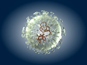
Hepatitis C virus. Computer artwork of hepatitis C virus particles (virions) in the human body. The virus consists of a core of RNA (ribonucleic acid) enclosed in a capsid

Background imageMicroorganisms Collection: EHEC E. coli bacteria, artwork C013 / 4621

EHEC E. coli bacteria, artwork C013 / 4621
EHEC E. coli bacteria. Computer artwork of a enterohaemorrhagic Escherichia coli (EHEC) bacteria in the human gut. E. coli bacteria are a normal part of the intestinal flora in humans

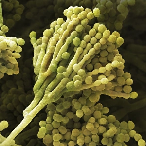
Background imageMicroorganisms Collection: Mould fungus, SEM

Mould fungus, SEM
Mould fungus (Penicillium expansum), coloured scanning electron micrograph (SEM). This fungus, also called blue mould, is a pathogen of plant material

Background imageMicroorganisms Collection: Staphylococcus aureus bacteria, SEM C013 / 4771

Staphylococcus aureus bacteria, SEM C013 / 4771
Staphylococcus aureus bacteria. Coloured scanning electron micrograph (SEM) of Staphylococcus aureus bacteria (yellow). S

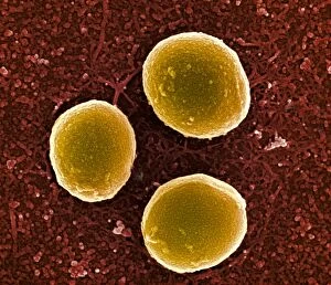
Background imageMicroorganisms Collection: Staphylococcus aureus bacteria, SEM C013 / 4770

Staphylococcus aureus bacteria, SEM C013 / 4770
Staphylococcus aureus bacteria. Coloured scanning electron micrograph (SEM) of Staphylococcus aureus bacteria (yellow). S

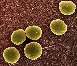
Background imageMicroorganisms Collection: Staphylococcus aureus bacteria, SEM C013 / 4769

Staphylococcus aureus bacteria, SEM C013 / 4769
Staphylococcus aureus bacteria. Coloured scanning electron micrograph (SEM) of Staphylococcus aureus bacteria (yellow). S

Background imageMicroorganisms Collection: Salmonella bacterium, artwork C013 / 4728

Salmonella bacterium, artwork C013 / 4728
Salmonella bacterium, computer artwork. Salmonella sp. bacteria are gram-negative rod-shaped bacteria that have flagella (hair-like structures) that they use for locomotion

Background imageMicroorganisms Collection: Salmonella bacteria, artwork C013 / 4727

Salmonella bacteria, artwork C013 / 4727
Salmonella bacteria, computer artwork. Salmonella sp. bacteria are gram-negative rod-shaped bacteria that have flagella (hair-like structures) that they use for locomotion

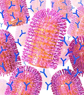
Background imageMicroorganisms Collection: Viral recognition by antibodies, artwork C013 / 4722

Viral recognition by antibodies, artwork C013 / 4722
Viral recognition by antibodies. Computer artwork of rabies (family Rhabdoviridae) virus particles (virions, pink) being identified by monoclonal antibodies (Y-shaped, blue)

Background imageMicroorganisms Collection: Rabies virus, artwork C013 / 4704

Rabies virus, artwork C013 / 4704
Rabies virus. Computer artwork of rabies (family Rhabdoviridae) virus particles (virions). The virus has a protein coat or capsid, which is made up of helically arranged subunits called capsomeres

Background imageMicroorganisms Collection: Rabies virus, artwork C013 / 4703

Rabies virus, artwork C013 / 4703
Rabies virus. Computer artwork of a rabies (family Rhabdoviridae) virus particle (virion). The virus has a protein coat or capsid, which is made up of helically arranged subunits called capsomeres

Background imageMicroorganisms Collection: Red Cyanobacteria (Cyanobacteria sp. ) forming scum on pool in freshwater marsh, Ceredigion

Red Cyanobacteria (Cyanobacteria sp. ) forming scum on pool in freshwater marsh, Ceredigion, Wales, june
Red Cyanobacteria (Cyanobacteria sp.) forming scum on pool in freshwater marsh, Ceredigion, Wales, june


All Professionally Made to Order for Quick Shipping

Microorganisms, the tiny wonders of life that exist all around us, are both fascinating and formidable. From the resilient water bear to the intricate structures captured in light micrographs, these microscopic creatures never cease to amaze. Infections spread by sneezing remind us of their potential impact on our health. The artwork depicting this transmission serves as a stark reminder of how easily these unseen organisms can wreak havoc within our bodies. The water bear, observed through scanning electron microscopy, showcases its remarkable resilience and adaptability. Its ability to survive extreme conditions has earned it the title of nature's toughest creature. Looking back at history, lithographs from 1906 reveal colonies of various bacteria that have plagued humanity for centuries. Haemophilus influenzae, Mycobacterium leprae, Micrococcus Gonorrhoea - each with their own unique characteristics and consequences for human health. Streptococcus pneumoniae takes center stage in another lithograph series; its bubble capsule highlighting its role in causing severe respiratory infections. These images serve as a testament to the ongoing battle between humans and disease-causing microorganisms throughout history. Spirochaetes Borrelia Recurrentis is responsible for Lyme disease - an illness that continues to affect countless individuals worldwide. A lithograph showcasing this spirillum highlights the importance of early detection and treatment when dealing with such infectious agents. Vibrio cholerae is yet another notorious bacterium depicted in a lithograph from 1906. This organism causes cholera outbreaks that have claimed countless lives over centuries past and present. Clostridium tetani reveals itself through spores in another captivating lithograph from 1906. This bacterium causes tetanus - a potentially deadly infection often associated with puncture wounds or contaminated objects. Lastly, Mycobacterium tuberculosis reminds us of one of humanity's oldest adversaries: tuberculosis. This ancient pathogen still poses significant challenges today despite medical advancements.