Fraud Blocker Skip to main content

Microorganism Collection (page 19)

Microorganisms, the tiny life forms that exist all around us, are truly fascinating

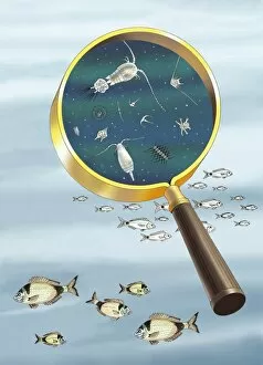
Background imageMicroorganism Collection: Plankton and fish, artwork C016 / 3448

Plankton and fish, artwork C016 / 3448
Plankton and fish. Artwork of plankton as viewed under a magnifying glass, with seabream (Diplodus vulgaris) fish

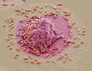
Background imageMicroorganism Collection: E. coli induced cell death, SEM C016 / 3078

E. coli induced cell death, SEM C016 / 3078
E. coli induced cell death. Coloured scanning electron micrograph (SEM) of a macrophage white blood cell (centre) that is being destroyed by toxins released by Escherichia coli (E)

Background imageMicroorganism Collection: E. coli induced cell death, SEM C016 / 3077

E. coli induced cell death, SEM C016 / 3077
E. coli induced cell death. Coloured scanning electron micrograph (SEM) of a macrophage white blood cell (centre) that is being destroyed by toxins released by Escherichia coli (E)

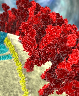
Background imageMicroorganism Collection: SARS coronavirus proteins, artwork C016 / 3056

SARS coronavirus proteins, artwork C016 / 3056
SARS coronavirus proteins. Computer model showing the spike proteins (red) of a SARS coronavirus particle (virion). SARS (severe acute respiratory syndrome)

Background imageMicroorganism Collection: SARS coronavirus, artwork C016 / 3055

SARS coronavirus, artwork C016 / 3055
SARS coronavirus proteins. Computer artwork of SARS coronavirus particles (virions). SARS (severe acute respiratory syndrome)

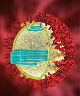
Background imageMicroorganism Collection: SARS coronavirus, artwork C016 / 3054

SARS coronavirus, artwork C016 / 3054
SARS coronavirus proteins. Cutaway computer model showing the protein structure of a SARS coronavirus particle (virion). SARS (severe acute respiratory syndrome)

Background imageMicroorganism Collection: SARS coronavirus, artwork C016 / 3053

SARS coronavirus, artwork C016 / 3053
SARS coronavirus proteins. Cutaway computer model showing the protein structure of a SARS coronavirus particle (virion). SARS (severe acute respiratory syndrome)

Background imageMicroorganism Collection: SARS coronavirus proteins, artwork C016 / 3052

SARS coronavirus proteins, artwork C016 / 3052
SARS coronavirus proteins. Computer model showing the proteins of a SARS coronavirus particle (virion). SARS (severe acute respiratory syndrome)

Background imageMicroorganism Collection: Vorticella protozoa, light micrograph C016 / 3042

Vorticella protozoa, light micrograph C016 / 3042
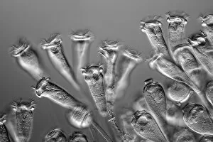
Vorticella protozoa. Differential interference contrast (DIC) light micrograph of Vorticella sp. ciliate protozoa. These single-celled organisms consist of a bell-shaped head

Background imageMicroorganism Collection: Vorticella protozoan, light micrograph C016 / 3043

Vorticella protozoan, light micrograph C016 / 3043
Vorticella protozoan. Differential interference contrast (DIC) light micrograph of a Vorticella sp. ciliate protozoan. This single-celled organism consists of a bell-shaped head

Background imageMicroorganism Collection: Vorticella protozoa, light micrograph C016 / 3041

Vorticella protozoa, light micrograph C016 / 3041
Vorticella protozoa. Differential interference contrast (DIC) light micrograph of Vorticella sp. ciliate protozoa. These single-celled organisms consist of a bell-shaped head

Background imageMicroorganism Collection: Vorticella protozoa, light micrograph C016 / 3040

Vorticella protozoa, light micrograph C016 / 3040
Vorticella protozoa. Differential interference contrast (DIC) light micrograph of Vorticella sp. ciliate protozoa. These single-celled organisms consist of a bell-shaped head

Background imageMicroorganism Collection: Satellite tobacco necrosis virus C014 / 0005

Satellite tobacco necrosis virus C014 / 0005
Satellite tobacco necrosis virus. Computer artwork showing the surface structure of a tobacco necrosis satellivirus particle (virion)

Background imageMicroorganism Collection: Hepatitis B virus particle C013 / 9983

Hepatitis B virus particle C013 / 9983
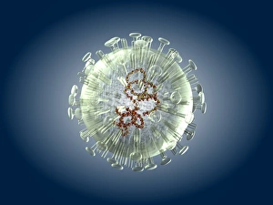
Hepatitis B virus particle. Computer artwork of the nucleocapsid of a hepatitis B virus (HBV) particle (virion). The capsid is a protein coat that encloses the viruss DNA (deoxyribonucleic acid)

Background imageMicroorganism Collection: Canine parvovirus particle C013 / 9966

Canine parvovirus particle C013 / 9966
Canine parvovirus particle. Computer artwork showing the structure of the outer protein coat (capsid) of a canine parvovirus type 2 particle (virion)

Background imageMicroorganism Collection: Salmonella bacteria, artwork C013 / 7809

Salmonella bacteria, artwork C013 / 7809
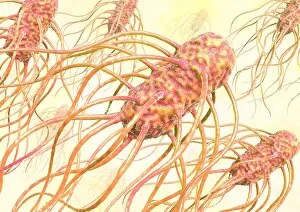
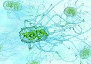
Salmonella bacteria, computer artwork. Salmonella sp. bacteria are gram-negative rod-shaped bacteria that have flagella (hair-like structures) that they use for locomotion

Background imageMicroorganism Collection: Salmonella bacteria, artwork C013 / 7808

Salmonella bacteria, artwork C013 / 7808
Salmonella bacteria, computer artwork. Salmonella sp. bacteria are gram-negative rod-shaped bacteria that have flagella (hair-like structures) that they use for locomotion

Background imageMicroorganism Collection: Bird flu virus particle, artwork C013 / 4650

Bird flu virus particle, artwork C013 / 4650
Bird flu virus particle. Computer artwork showing the internal and external structures of an influenza (flu) A subtype H5N1 (bird flu) virus particle (virion)

Background imageMicroorganism Collection: Ross River virus particle, artwork C013 / 4641

Ross River virus particle, artwork C013 / 4641
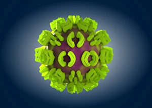
Ross River virus particle. Computer artwork showing the outer structure of a Ross River virus (RRV) particle (virion). RRV is a small encapsulated single-strand RNA alphavirus endemic to Australia

Background imageMicroorganism Collection: Marburg virus, artwork C013 / 4633

Marburg virus, artwork C013 / 4633
Marburg virus. Computer artwork of Marburg virus particles (virions). This tubular RNA (ribonucleic acid, coloured spheres) virus causes Marburg haemorrhagic fever in humans and non-human primates

Background imageMicroorganism Collection: Hepatitis C virus, artwork

Hepatitis C virus, artwork
Hepatitis C virus. Computer artwork of hepatitis C virus particles (virions) in the human body. The virus consists of a core of RNA (ribonucleic acid) enclosed in a capsid

Background imageMicroorganism Collection: EHEC E. coli bacteria, artwork C013 / 4621

EHEC E. coli bacteria, artwork C013 / 4621
EHEC E. coli bacteria. Computer artwork of a enterohaemorrhagic Escherichia coli (EHEC) bacteria in the human gut. E. coli bacteria are a normal part of the intestinal flora in humans

Background imageMicroorganism Collection: Mould fungus, SEM

Mould fungus, SEM
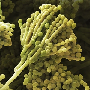
Mould fungus (Penicillium expansum), coloured scanning electron micrograph (SEM). This fungus, also called blue mould, is a pathogen of plant material

Background imageMicroorganism Collection: Staphylococcus aureus bacteria, SEM C013 / 4771

Staphylococcus aureus bacteria, SEM C013 / 4771
Staphylococcus aureus bacteria. Coloured scanning electron micrograph (SEM) of Staphylococcus aureus bacteria (yellow). S

Background imageMicroorganism Collection: Staphylococcus aureus bacteria, SEM C013 / 4770

Staphylococcus aureus bacteria, SEM C013 / 4770
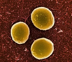
Staphylococcus aureus bacteria. Coloured scanning electron micrograph (SEM) of Staphylococcus aureus bacteria (yellow). S

Background imageMicroorganism Collection: Staphylococcus aureus bacteria, SEM C013 / 4769

Staphylococcus aureus bacteria, SEM C013 / 4769
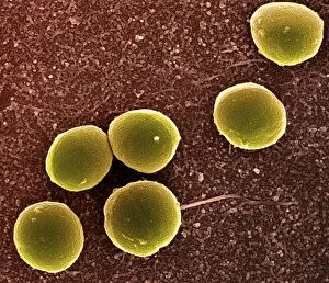
Staphylococcus aureus bacteria. Coloured scanning electron micrograph (SEM) of Staphylococcus aureus bacteria (yellow). S

Background imageMicroorganism Collection: Salmonella bacterium, artwork C013 / 4728

Salmonella bacterium, artwork C013 / 4728
Salmonella bacterium, computer artwork. Salmonella sp. bacteria are gram-negative rod-shaped bacteria that have flagella (hair-like structures) that they use for locomotion

Background imageMicroorganism Collection: Salmonella bacteria, artwork C013 / 4727

Salmonella bacteria, artwork C013 / 4727
Salmonella bacteria, computer artwork. Salmonella sp. bacteria are gram-negative rod-shaped bacteria that have flagella (hair-like structures) that they use for locomotion

Background imageMicroorganism Collection: Viral recognition by antibodies, artwork C013 / 4722

Viral recognition by antibodies, artwork C013 / 4722
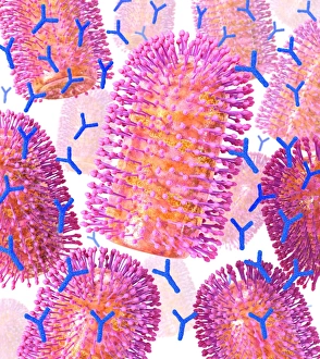
Viral recognition by antibodies. Computer artwork of rabies (family Rhabdoviridae) virus particles (virions, pink) being identified by monoclonal antibodies (Y-shaped, blue)

Background imageMicroorganism Collection: Rabies virus, artwork C013 / 4704

Rabies virus, artwork C013 / 4704
Rabies virus. Computer artwork of rabies (family Rhabdoviridae) virus particles (virions). The virus has a protein coat or capsid, which is made up of helically arranged subunits called capsomeres

Background imageMicroorganism Collection: Rabies virus, artwork C013 / 4703

Rabies virus, artwork C013 / 4703
Rabies virus. Computer artwork of a rabies (family Rhabdoviridae) virus particle (virion). The virus has a protein coat or capsid, which is made up of helically arranged subunits called capsomeres

Background imageMicroorganism Collection: Red Cyanobacteria (Cyanobacteria sp. ) forming scum on pool in freshwater marsh, Ceredigion

Red Cyanobacteria (Cyanobacteria sp. ) forming scum on pool in freshwater marsh, Ceredigion, Wales, june
Red Cyanobacteria (Cyanobacteria sp.) forming scum on pool in freshwater marsh, Ceredigion, Wales, june


All Professionally Made to Order for Quick Shipping

Microorganisms, the tiny life forms that exist all around us, are truly fascinating. From HeLa cells to water bears and viruses like MRSA and RSV, these microscopic entities play a significant role in our lives. In the world of science, HeLa cells have been instrumental in numerous breakthroughs. Captured under a light micrograph (C017 / 8299), their intricate structures reveal the complexity of human biology. Another captivating image shows neutrophils engulfing MRSA bacteria, as seen through a scanning electron microscope (C018 / 8596). This powerful defense mechanism showcases how our immune system fights against harmful invaders. Water bears, also known as tardigrades, are remarkable creatures that can survive extreme conditions. A light micrograph (C016 / 8581) captures their unique appearance with their stout bodies and claw-like limbs. Equally intriguing is another image taken using a scanning electron microscope (SEM C016 / 9084), which reveals even more details about these resilient organisms. Viruses such as respiratory syncytial virus and flu virus particles have caused widespread infections throughout history. While one can only be observed under a microscope (microscopic view of human respiratory syncytial virus), the other is depicted through artwork showcasing its structure (flu virus particle, artwork F008 / 3245). These images remind us of the importance of vaccinations in preventing viral outbreaks. The spread of infections by sneezing is illustrated through an artistic representation (artwork C013 / 5949). It serves as a reminder to practice good hygiene to protect ourselves and others from contagious diseases. Chlamydia may be small but has significant consequences for those affected by it. A microscopic view highlights its presence within host cells—a stark reminder of the need for awareness and prevention efforts against sexually transmitted infections. Looking back at historical lithographs from 1906 brings attention to colonies formed by Haemophilus influenzae and Mycobacterium leprae.