Fraud Blocker Skip to main content

Micrograph Collection (page 8)

Exploring the intricate world of micrographs: From synapse nerve junctions captured through TEM to the mesmerizing beauty of copper and magnesium sulphate under LM

Background imageMicrograph Collection: Picture No. 10873703

Picture No. 10873703
Sleeping Sickness Parasite in red blood cells (Trypanosoma sp.) Date:

Background imageMicrograph Collection: Picture No. 10873596

Picture No. 10873596
Human Cochlea Date:

Background imageMicrograph Collection: Picture No. 10877011

Picture No. 10877011
Scanning Electron Micrograph (SEM): Frog tongue - taste receptors papillae Date:

Background imageMicrograph Collection: Picture No. 10877020

Picture No. 10877020
Scanning Electron micrograph (SEM): Open Stoma of a plant leaf; Date:

Background imageMicrograph Collection: Picture No. 10877013

Picture No. 10877013
Scanning Electron Micrograph (SEM); Gecko Claw: Date:

Background imageMicrograph Collection: Picture No. 10877021

Picture No. 10877021
Scanning Electron micrograph (SEM): Open Stoma of a plant leaf; Date:

Background imageMicrograph Collection: Picture No. 10877014

Picture No. 10877014
Scanning Electron Micrograph (SEM): Gecko foot hairs Date:

Background imageMicrograph Collection: Picture No. 10877012

Picture No. 10877012
Scanning Electron Micrograph (SEM): human intestine showing villi Date:

Background imageMicrograph Collection: Picture No. 10877008

Picture No. 10877008
Scanning Electron Micrograph (SEM) of a resin cast of blood vessels in the intestines; Date:

Background imageMicrograph Collection: Picture No. 10877005

Picture No. 10877005
Scanning Electron Micrograph (SEM): Human hair - Asian Date:

Background imageMicrograph Collection: Picture No. 10877007

Picture No. 10877007
Scanning Electron Micrograph (SEM): Human Hairs Date:

Background imageMicrograph Collection: Picture No. 10876998

Picture No. 10876998
Scanning Electron Micrograph (SEM): Human Skin Date:

Background imageMicrograph Collection: Picture No. 10877000

Picture No. 10877000
Scanning Electron Micrograph (SEM): Human Skin Date:

Background imageMicrograph Collection: Picture No. 10877004

Picture No. 10877004
Scanning Electron Micrograph (SEM): Human hair - European Date:

Background imageMicrograph Collection: Picture No. 10877006

Picture No. 10877006
Scanning Electron Micrograph (SEM): Human pubic hair Date:

Background imageMicrograph Collection: Picture No. 10877003

Picture No. 10877003
Scanning Electron Micrograph (SEM): Human hair - African Date:

Background imageMicrograph Collection: Picture No. 10876996

Picture No. 10876996
Scanning Electron Micrograph (SEM): Human Retina showing photoreceptors rod and cone cells Date:

Background imageMicrograph Collection: Picture No. 10877001

Picture No. 10877001
Scanning Electron Micrograph (SEM): Human Skin Date:

Background imageMicrograph Collection: Picture No. 10876999

Picture No. 10876999
Scanning Electron Micrograph (SEM): Human Skin Date:

Background imageMicrograph Collection: Picture No. 10876991

Picture No. 10876991
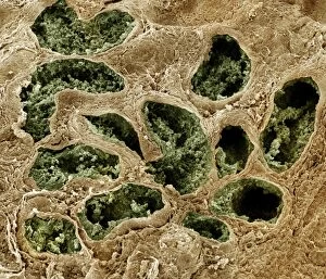
Scanning Electron Micrograph (SEM): Human testis tubes - Sperm cells forming in seminal tubes of testis Date:

Background imageMicrograph Collection: Picture No. 10876993

Picture No. 10876993
Scanning Electron Micrograph (SEM): Human Retina showing photoreceptors rod and cone cells Date:

Background imageMicrograph Collection: Picture No. 10876995

Picture No. 10876995
Scanning Electron Micrograph (SEM): Human Retina showing photoreceptors rod and cone cells Date:

Background imageMicrograph Collection: Picture No. 10876989

Picture No. 10876989
Scanning Electron Micrograph (SEM): Human Fallopian tube Date:

Background imageMicrograph Collection: Picture No. 10876994

Picture No. 10876994
Scanning Electron Micrograph (SEM): Human Retina showing photoreceptors rod and cone cells Date:

Background imageMicrograph Collection: Picture No. 10876992

Picture No. 10876992
Scanning electron micrograph (SEM) of Kidney glomerulus Date:

Background imageMicrograph Collection: Picture No. 10876988

Picture No. 10876988
Scanning Electron Micrograph (SEM): Early human embryo, one cell removed for genetic analysis Date:

Background imageMicrograph Collection: Picture No. 10876986

Picture No. 10876986
Scanning Electron Micrograph (SEM): Human Red Blood Cells Date:

Background imageMicrograph Collection: Picture No. 10875840

Picture No. 10875840
Light Micrograph of Atherosclerotic Plaque and Obstructed Lumen of Human Aorta Date:

Background imageMicrograph Collection: Picture No. 10875844

Picture No. 10875844
Light Micrograph of Atherosclerotic Plaque and Obstructed Lumen of Human Aorta Date:

Background imageMicrograph Collection: Picture No. 10875843

Picture No. 10875843
Light Micrograph of Atherosclerotic Plaque and Obstructed Lumen of Human Aorta Date:

Background imageMicrograph Collection: Picture No. 10875841

Picture No. 10875841
Light Micrograph of Atherosclerotic Aorta Vessel Date:

Background imageMicrograph Collection: Picture No. 10875839

Picture No. 10875839
Light Micrograph of Atherosclerotic Plaque and Obstructed Lumen of Human Aorta Date:

Background imageMicrograph Collection: Picture No. 10875842

Picture No. 10875842
Light Micrograph of Atherosclerotic Plaque and Obstructed Lumen of Human Aorta Date:

Background imageMicrograph Collection: Picture No. 10875838

Picture No. 10875838
Light Micrograph of Atherosclerotic Plaque and Obstructed Lumen of Human Aorta Date:

Background imageMicrograph Collection: Picture No. 10875837

Picture No. 10875837
Light Micrograph of Atherosclerotic Plaque and Obstructed Lumen of Human Aorta Date:

Background imageMicrograph Collection: Picture No. 10875836

Picture No. 10875836
Light Micrograph of Atherosclerotic Plaque and Obstructed Lumen of Human Aorta Date:

Background imageMicrograph Collection: Picture No. 10875812

Picture No. 10875812
Light Micrograph of Cavitated Mouse Blastocyst Date:

Background imageMicrograph Collection: Picture No. 10875811

Picture No. 10875811
Light Micrograph of Mouse Blastocyst Date:

Background imageMicrograph Collection: Picture No. 10875805

Picture No. 10875805
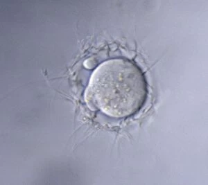
Light Micrograph of Mouse Ovum being fertilized Date:

Background imageMicrograph Collection: Picture No. 10875802

Picture No. 10875802
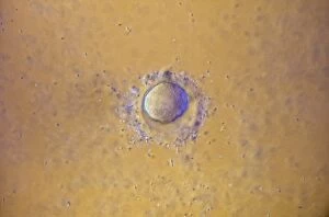
Light Micrograph of human Ovum surrounded by Sperm - being fertilized Date:

Background imageMicrograph Collection: Picture No. 10875806

Picture No. 10875806
Light Micrograph of Mouse Ovum being fertilized Date:

Background imageMicrograph Collection: Picture No. 10875800

Picture No. 10875800
Light Micrograph of Human Sperm; Date:


All Professionally Made to Order for Quick Shipping

Exploring the intricate world of micrographs: From synapse nerve junctions captured through TEM to the mesmerizing beauty of copper and magnesium sulphate under LM. Witness the delicate structure of Discosphaera tubifera, a coccolithophore, or marvel at the dangerous Crysotile asbestos fibers. Delve into the inner workings of a liver cell or encounter the notorious Cimex lectularius, better known as bed bugs. Admire the ethereal dandelion fruiting head, Taraxacum officinale, or study Simulium damnosum, a Simulian blackfly responsible for river blindness. Uncover E. coli bacteria's microscopic realm or be amazed by snail teeth that defy their size. Behold a stunning silicon crystal in all its glory through light micrography and observe C. elegans worms navigating their environment with grace. These glimpses into unseen worlds remind us of nature's complexity and endless wonders.