
Microglial Collection
Microglial cells, also known as microglia, are a type of glial cell found in the central nervous system


All Professionally Made to Order for Quick Shipping
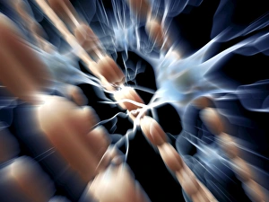
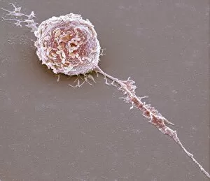
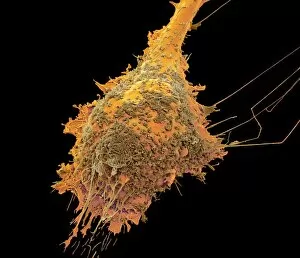
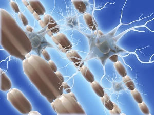
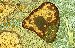
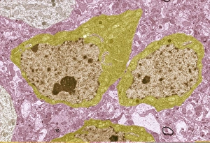
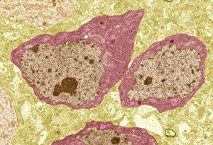
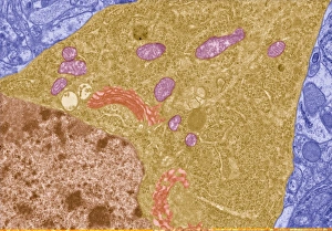
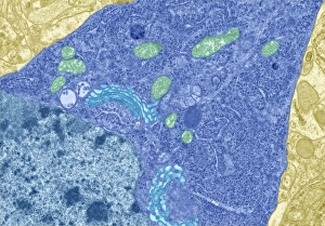
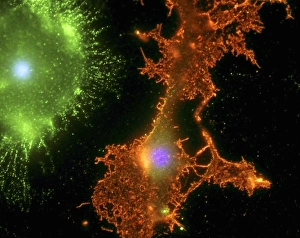
Microglial cells, also known as microglia, are a type of glial cell found in the central nervous system. They play a crucial role in maintaining brain health and function. One fascinating aspect of microglia is their ability to interact with myelin sheaths, which are protective coverings that surround nerve fibers. These sheaths help facilitate efficient transmission of electrical signals between neurons. In artwork C014 / 2646, we can see the intricate network of myelin sheaths intertwined with glial cells, including microglia. This depiction showcases the close relationship between these structures and highlights their importance in supporting neural communication. SEM images such as C016 / 9115, C016 / 9119, C016 / 9117, C016 / 9109, C016 / 9114, C016 / 9113, and C016 / 9118 provide us with a closer look at individual microglial white blood cells. These microscopic warriors tirelessly patrol the brain for any signs of damage or infection. The SEM images (C016/9111) and (C016/9112) further emphasize the abundance and diversity of these remarkable immune cells within our central nervous system. Studying microglia is essential for understanding various neurological disorders like Alzheimer's disease or multiple sclerosis where dysfunction or dysregulation of these cells may contribute to pathology. In image (C014/2647), we witness another captivating portrayal depicting myelin sheaths alongside glial cells. This artwork serves as a reminder that while we marvel at scientific advancements like scanning electron microscopy (SEM), artistic interpretations continue to inspire curiosity about our complex biological systems.