
Microglia Collection
Microglia, the unsung heroes of our central nervous system, play a crucial role in maintaining its health and functionality


All Professionally Made to Order for Quick Shipping
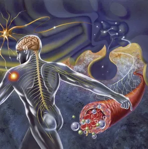
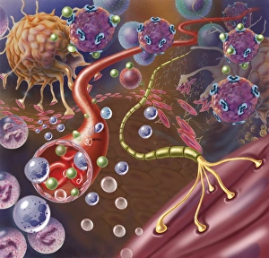
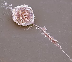
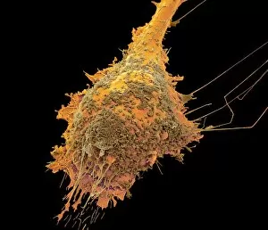
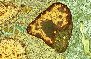
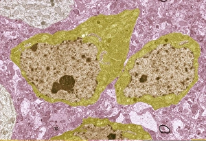
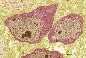
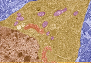
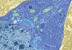
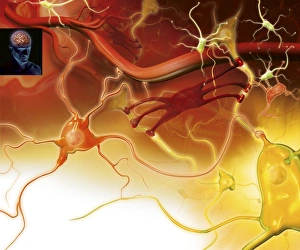
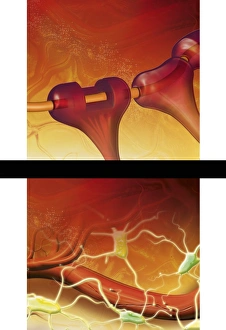
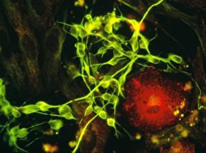
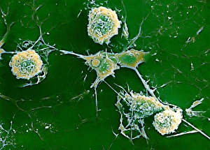
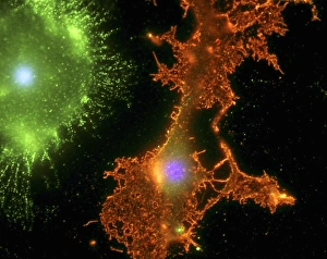
Microglia, the unsung heroes of our central nervous system, play a crucial role in maintaining its health and functionality. These remarkable cells have been at the forefront of research on neurological disorders like multiple sclerosis. In an astonishing SEM image, we witness the intricate network of microglial white blood cells within our hypothalamus. This region receives nerve impulses from all corners of our body, ensuring seamless communication between our brain and organs. Zooming in closer, we observe a nerve with its protective myelin sheath connecting with a muscle. Microglia stand guard nearby, ready to spring into action if any threat arises. Their presence is vital for preserving the integrity and function of this delicate connection. Examining individual microglial white blood cells under high magnification reveals their fascinating structure and complexity. SEM images showcase these tiny warriors in various stages: C016 / 9115, C016 / 9119, C016 / 9117 - each one unique yet united by their mission to protect us. These microscopic defenders tirelessly patrol our brain tissue day and night. In SEM images such as C016 / 9109 or C016 / 9114, we witness their tireless dedication as they survey their surroundings for signs of trouble. Together as a team (as seen in SEM image C016 / 9113), microglia form an army that stands ready to combat any potential threats lurking within our central nervous system. Their vigilance ensures that harmful substances are swiftly eliminated while promoting healing processes when needed most. SEM images like C016 / 9118 or those featuring clusters such as C016 / 9111 and C016/9112 highlight the sheer numbers at which these incredible cells operate within us – always prepared to defend against invaders or repair damaged neural pathways. As researchers continue unraveling the mysteries surrounding microglia's functions and capabilities, it becomes increasingly evident just how essential they are for our neurological well-being.