
Microbe Collection
"Unveiling the Invisible: Exploring the Microbial World" In a realm unseen by the naked eye, lies a fascinating universe teeming with life


All Professionally Made to Order for Quick Shipping
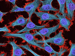
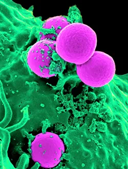
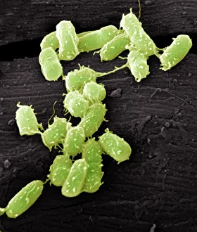
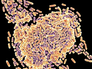
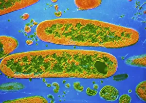
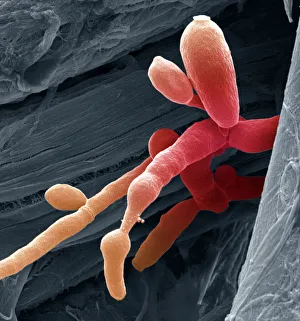
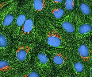
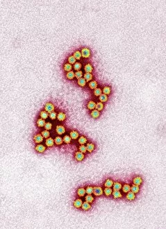
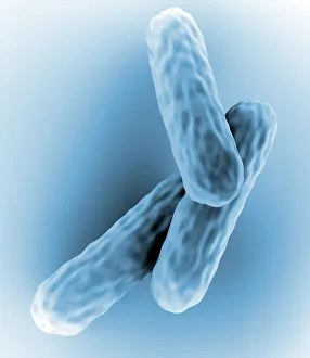
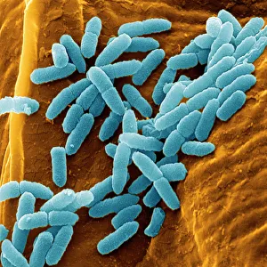
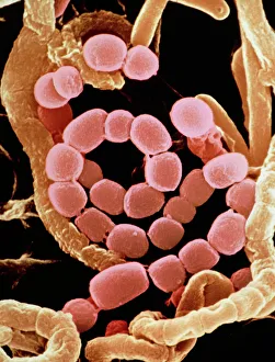
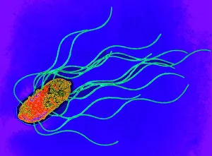
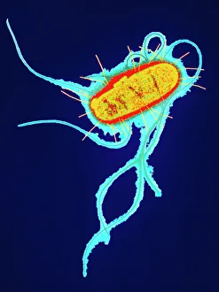
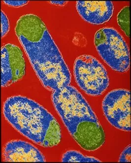
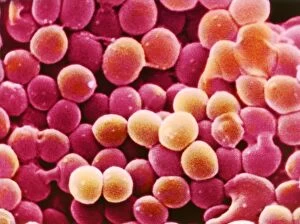
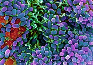
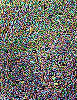
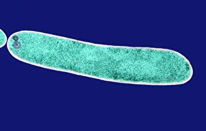
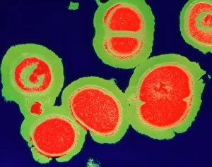
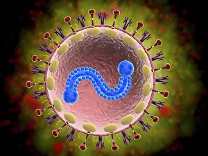
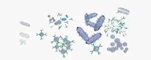
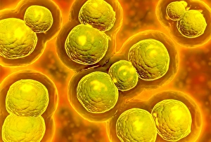
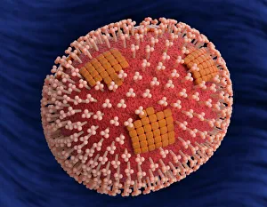
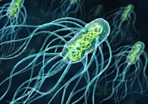
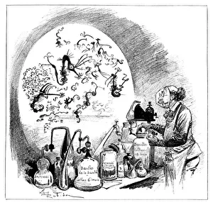
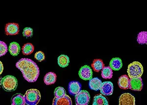
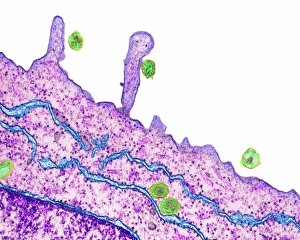
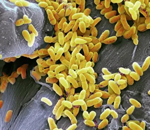
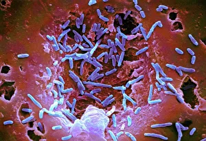
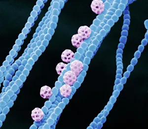
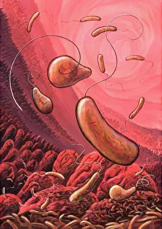
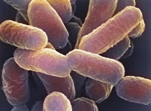
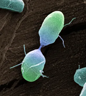
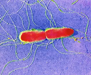
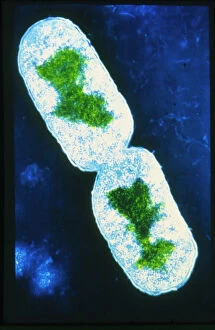
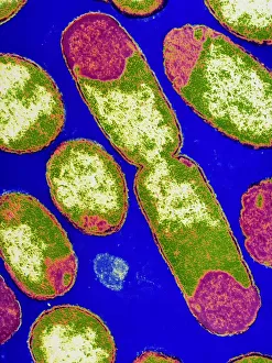
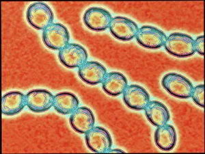
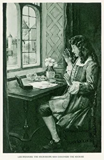
"Unveiling the Invisible: Exploring the Microbial World" In a realm unseen by the naked eye, lies a fascinating universe teeming with life. Meet the microbe, an extraordinary entity that encompasses various organisms and shapes our understanding of biology. HeLa cells, immortalized and widely used in research, have revolutionized medical science as they continue to unravel mysteries within our own bodies. Captured under a light microscope (C017 / 8299), their intricate structures reveal secrets waiting to be discovered. Witness the battle between neutrophils and MRSA as these microscopic warriors engage in a relentless struggle for survival. In an astonishing scanning electron microscope image (C018 / 8596), observe how these immune cells engulf harmful bacteria, showcasing nature's defense mechanisms at work. Eagerly multiplying like tiny soldiers on a mission, E. coli bacteria emerge into view through another SEM image. Their presence reminds us of both their beneficial role in digestion and potential harm when found in contaminated food or water sources. Salmonella bacteria take center stage next; their distinctive features magnified by yet another SEM image. These notorious culprits behind foodborne illnesses serve as reminders of proper hygiene practices necessary for safeguarding public health. Behold Yersinia pestis bacteria captured in vibrant colors through transmission electron microscopy (TEM). This captivating visual representation highlights the infamous pathogen responsible for devastating outbreaks such as the Black Death throughout history. Delving deeper into this microbial world reveals Candida fungus thriving amidst its surroundings. A striking SEM image showcases its filamentous structure – reminding us of its ability to cause infections if given favorable conditions. Returning to HeLa cells once more (light micrograph C017 / 8298), we are reminded of Henrietta Lacks' invaluable contribution to medical research - her immortal cell line continues to pave new paths towards scientific breakthroughs even decades after her passing.