Fraud Blocker Skip to main content

Metatarsus Collection

Metatarsus, also known as the metatarsal bones, is a crucial part of the human skeletal system

Background imageMetatarsus Collection: Diagram of the bones of the right leg and hip

Diagram of the bones of the right leg and hip
Diagram of the bones of the right leg, showing the joint with the pelvis at the hip

Background imageMetatarsus Collection: Human foot anatomy showing skin, veins, arteries, muscles and bones

Human foot anatomy showing skin, veins, arteries, muscles and bones
Human foot anatomy showing skin, veins, arteries, muscles, and bones, cutaway view

Background imageMetatarsus Collection: Outer ankle ligaments, artwork C013 / 4452

Outer ankle ligaments, artwork C013 / 4452
Outer ankle ligaments. Computer artwork of the bones and ligaments (white) of the right foot and ankle seen from the side

Background imageMetatarsus Collection: Inner ankle ligaments, artwork C013 / 4451

Inner ankle ligaments, artwork C013 / 4451
Inner ankle ligaments. Computer artwork of the bones and ligaments (white) of the right foot and ankle seen from the side

Background imageMetatarsus Collection: Tibia and metatarsus of the extinct Rodrigues solitaire, Pezophaps solitaria

Tibia and metatarsus of the extinct Rodrigues solitaire, Pezophaps solitaria
FLO4612197 Tibia and metatarsus of the extinct Rodrigues solitaire, Pezophaps solitaria, in the Parisian Collection and Andersonian Collection

Background imageMetatarsus Collection: Metatarsus and toes of the dodo, Raphus cucullatus 1-5, foot of the yellow-footed green pigeon

Metatarsus and toes of the dodo, Raphus cucullatus 1-5, foot of the yellow-footed green pigeon, Treron chlorigaster 6
FLO4612178 Metatarsus and toes of the dodo, Raphus cucullatus 1-5, foot of the yellow-footed green pigeon, Treron chlorigaster 6, stock dove, Columba oenas 7 and partridge pigeon

Background imageMetatarsus Collection: Healthy ankle joint, X-ray

Healthy ankle joint, X-ray
Healthy ankle joint. Coloured profile X-ray of the left ankle of a 21 year old patient. Strapping around the ankle is visible on this X-ray

Background imageMetatarsus Collection: Tibia and metatarsus of the extinct Rodrigues

Tibia and metatarsus of the extinct Rodrigues solitaire, Pezophaps solitaria, in the Parisian Collection and Andersonian Collection

Background imageMetatarsus Collection: Metatarsus and toes of the dodo and various pigeons

Metatarsus and toes of the dodo and various pigeons
Metatarsus and toes of the dodo, Raphus cucullatus 1-5, foot of the yellow-footed green pigeon, Treron chlorigaster 6, stock dove, Columba oenas 7 and partridge pigeon, Geophaps smithii 8

Background imageMetatarsus Collection: Metatarsus bones of the dodo, crowned pigeon

Metatarsus bones of the dodo, crowned pigeon
Metatarsus bones of the dodo, Raphus cucullatus 1-10, crowned pigeon, Goura coronata 12-17, tooth-billed pigeon, Didunculus strigirostris 18, 19 and other birds

Background imageMetatarsus Collection: Human skeletal system, front view

Human skeletal system, front view

Background imageMetatarsus Collection: Anterior view of human skeletal system, with labels

Anterior view of human skeletal system, with labels

Background imageMetatarsus Collection: Skeleton of an Eagle, after Milne-Edwards (engraving)

Skeleton of an Eagle, after Milne-Edwards (engraving)
966625 Skeleton of an Eagle, after Milne-Edwards (engraving) by English School, (19th century); Private Collection; (add.info.: Skeleton of an Eagle, after Milne-Edwards)

Background imageMetatarsus Collection: Male human skeleton in dynamic posture, front view

Male human skeleton in dynamic posture, front view

Background imageMetatarsus Collection: Front view of human skeleton waving goodbye

Front view of human skeleton waving goodbye, isolated on white background

Background imageMetatarsus Collection: Male human skeleton in dynamic posture, rear view

Male human skeleton in dynamic posture, rear view

Background imageMetatarsus Collection: Anterior view of human skeletal system

Anterior view of human skeletal system

Background imageMetatarsus Collection: Front view of human skeleton meditating

Front view of human skeleton meditating, isolated on white background

Background imageMetatarsus Collection: Human skeleton praying on his knees

Human skeleton praying on his knees, isolated on white background

Background imageMetatarsus Collection: Illustration of webbed foot of water bird

Illustration of webbed foot of water bird

Background imageMetatarsus Collection: Conceptual image of human legs and feet with nervous system

Conceptual image of human legs and feet with nervous system

Background imageMetatarsus Collection: Conceptual image of bones in human legs and feet

Conceptual image of bones in human legs and feet

Background imageMetatarsus Collection: Front view of human skeletal system

Front view of human skeletal system

Background imageMetatarsus Collection: Front view of human skeleton meditation

Front view of human skeleton meditation, isolated on white background

Background imageMetatarsus Collection: X-ray view of human foot

X-ray view of human foot

Background imageMetatarsus Collection: Conceptual image of pain in human foot

Conceptual image of pain in human foot

Background imageMetatarsus Collection: Bones in the human foot

Bones in the human foot

Background imageMetatarsus Collection: Conceptual image of gout in the big toe

Conceptual image of gout in the big toe. Gout is a form of inflammatory arthritis that causes sudden, severe pain, swelling and tenderness of joints

Background imageMetatarsus Collection: Medical illustration of human leg muscles, bones and joints

Medical illustration of human leg muscles, bones and joints

Background imageMetatarsus Collection: Dorsal anatomy of the human foot

Dorsal anatomy of the human foot

Background imageMetatarsus Collection: Front view of human skeleton

Front view of human skeleton, isolated on white background

Background imageMetatarsus Collection: Front view of human skeleton running

Front view of human skeleton running, isolated on white background

Background imageMetatarsus Collection: 3D rendering of human skeletal system, side view

3D rendering of human skeletal system, side view

Background imageMetatarsus Collection: Human foot with nervous system, lymphatic system and circulatory system

Human foot with nervous system, lymphatic system and circulatory system

Background imageMetatarsus Collection: Anterior compartment anatomy of left leg muscles and tendons

Anterior compartment anatomy of left leg muscles and tendons

Background imageMetatarsus Collection: Three dimensional view of human leg and feet bones

Three dimensional view of human leg and feet bones

Background imageMetatarsus Collection: 3D rendering of human skeletal system at different angles

3D rendering of human skeletal system at different angles

Background imageMetatarsus Collection: X-ray view of pain in human foot

X-ray view of pain in human foot

Background imageMetatarsus Collection: Healthy ankle joint, X-ray F006 / 9129

Healthy ankle joint, X-ray F006 / 9129
Healthy ankle joint. Coloured profile X-ray of the left ankle of a 21 year old patient. Strapping around the ankle is visible on this X-ray

Background imageMetatarsus Collection: Healthy ankle joint, X-ray F006 / 9127

Healthy ankle joint, X-ray F006 / 9127
Healthy ankle joint. Coloured frontal X-ray of the left ankle of a 21 year old patient. Strapping around the ankle is visible on this X-ray

Background imageMetatarsus Collection: Healthy ankle joint, X-ray F006 / 9126

Healthy ankle joint, X-ray F006 / 9126
Healthy ankle joint. Coloured frontal X-ray of the left ankle of a 21 year old patient. Strapping around the ankle is visible on this X-ray

Background imageMetatarsus Collection: Human foot, historical anatomical model

Human foot, historical anatomical model
Human foot, anatomical model. 1965 model showing the skeletal structure of the left human foot and ankle

Background imageMetatarsus Collection: Foot muscles, artwork C013 / 4487

Foot muscles, artwork C013 / 4487
Foot muscles. Computer artwork of a view looking down on some of the muscles and tendons (red and white) of the upper surface of the foot

Background imageMetatarsus Collection: Foot muscles, artwork C013 / 4486

Foot muscles, artwork C013 / 4486
Foot muscles. Computer artwork of an oblique anterior view of some of the muscles and tendons (red and white) of the upper surface of the foot

Background imageMetatarsus Collection: Foot muscles, artwork C013 / 4485

Foot muscles, artwork C013 / 4485
Foot muscles. Computer artwork of a view from below of some of the muscles and tendons (red and white) of the sole of the foot

Background imageMetatarsus Collection: Foot muscles, artwork C013 / 4483

Foot muscles, artwork C013 / 4483
Foot muscles. Computer artwork of an oblique view from below and behind of some of the muscles and tendons (red and white) of the sole of the foot

Background imageMetatarsus Collection: Foot muscles, artwork C013 / 4478

Foot muscles, artwork C013 / 4478
Foot muscles. Computer artwork of a view from below of some of the muscles and tendons (red and white) of the sole of the foot

Background imageMetatarsus Collection: Foot muscles, artwork C013 / 4477

Foot muscles, artwork C013 / 4477
Foot muscles. Computer artwork of a view from below of some of the muscles and tendons (red and white) of the sole of the foot


All Professionally Made to Order for Quick Shipping

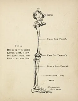
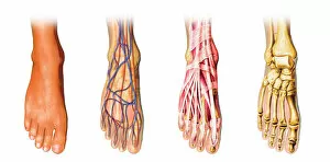
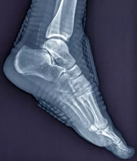
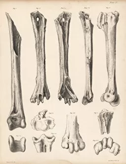
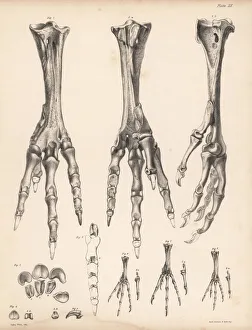
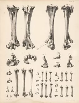
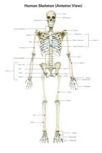
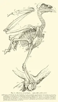
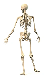
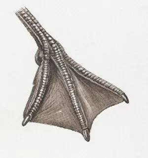
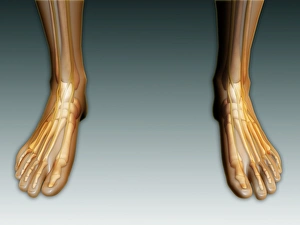
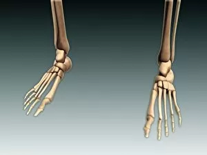
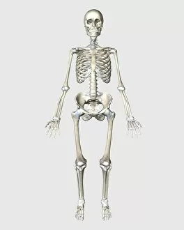
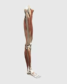
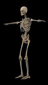
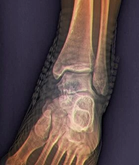
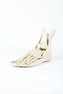
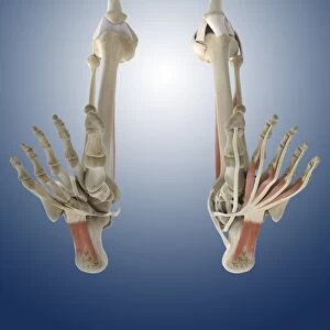
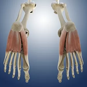
Metatarsus, also known as the metatarsal bones, is a crucial part of the human skeletal system. Located in the foot, it consists of five long bones that connect to the toes. These bones play a vital role in supporting our body weight and facilitating movement. When we look at a diagram of the right leg and hip, we can clearly see how the metatarsus fits into this intricate structure. It connects to the tibia and fibula through various ligaments, ensuring stability and flexibility during activities such as walking or running. In an anatomical illustration of the human foot, we can observe not only the skin but also veins, arteries, muscles, and bones. The metatarsus stands out prominently as one of these essential components. Its position between the ankle joint and phalanges demonstrates its significance in maintaining balance and absorbing shock while engaging in physical activities. Artworks depicting outer ankle ligaments (C013 / 4452) and inner ankle ligaments (C013 / 4451) shed light on how these structures support proper alignment within our feet. A healthy ankle joint X-ray showcases how well-preserved metatarsals contribute to overall foot health. Interestingly enough, even extinct species like Rodrigues' dodo had their own version bone structures. Comparing them with those found in various pigeons reveals evolutionary adaptations for different lifestyles. The human skeletal system's front view provides us with a comprehensive understanding of where exactly each bone lies within our bodies – including every individual metatarsal bone that forms part of this complex framework. An engraving showcasing an eagle's skeleton reminds us that birds too possess their own unique set of metatarsals adapted for flight purposes. This further emphasizes just how versatile these bones are across different species. Finally, observing a male human skeleton dynamically posed from a frontal perspective highlights not only his musculature but also underscores how interconnected all parts are – including the metatarsus.