
Membranes Collection
"Exploring the Intricate World of Membranes: From Chloroplasts to Bacterial Meningitis" Delving into the depths of cellular structures


All Professionally Made to Order for Quick Shipping
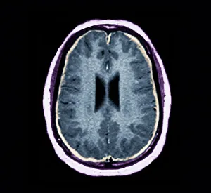
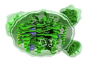
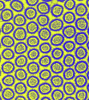
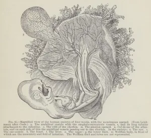
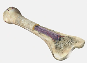
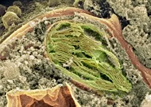
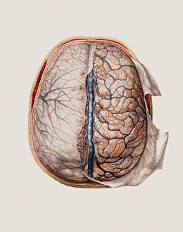
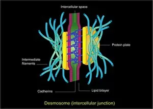
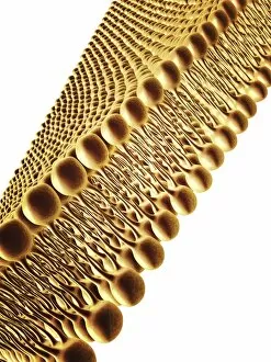
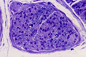
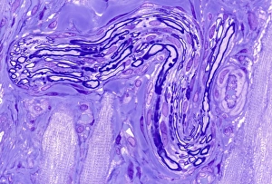
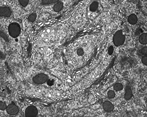
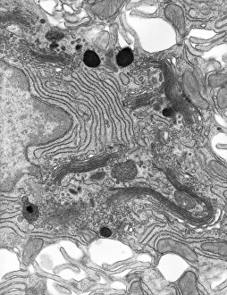
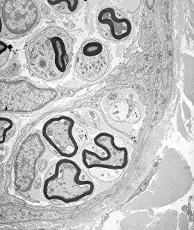
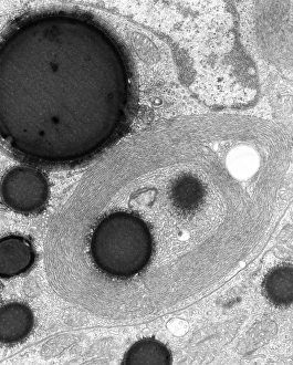
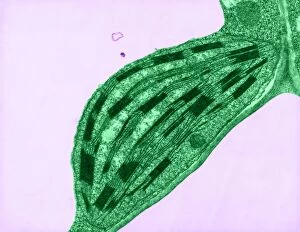
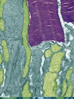
"Exploring the Intricate World of Membranes: From Chloroplasts to Bacterial Meningitis" Delving into the depths of cellular structures, we uncover the mesmerizing beauty of chloroplast membranes through intricate artwork. Shedding light on a medical mystery, an MRI scan reveals the devastating effects of bacterial meningitis on brain membranes. Peering into the microscopic realm, a TEM image showcases the delicate and vital role played by intestinal microvilli in nutrient absorption. Safeguarding our most precious organ, brain meninges shield and protect our cognitive powerhouse with unwavering dedication. Unveiling energy powerhouses within cells, SEM imaging captures stunning details of mitochondria's complex membrane structure. Witnessing an artistic representation of "Invasion de la gorge et du larynx par les fausses membranes, " we are reminded of the harrowing impact that diphtheria can have on throat and larynx membranes (colour litho). Journeying back to life's earliest stages, an engraving offers a magnified view of a four-week-old human embryo with its protective membranes opened wide. Exploring bone anatomy at its core, a cross-sectional diagram highlights femur's osteon network alongside veins and marrow while emphasizing membrane significance. Revealing nature's architectural marvels once more, SEM imagery unveils intricate details within chloroplasts' membranous structures responsible for photosynthesis. Appreciating artistry in scientific depictions, F007 / 1477 artwork portrays cell membrane lipid bilayer intricacies - showcasing their importance in cellular function. Returning to safeguard our brains yet again; let us marvel at brain meninges' resilience as they continue their tireless duty day after day. Revisiting mitochondria through SEM imaging allows us to witness their awe-inspiring complexity once more – reminding us of their crucial role in cellular energy production.