
Medical Scan Collection
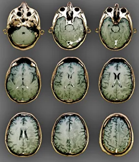
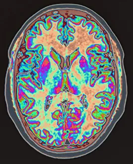
"Exploring the Intricacies of Medical Scan: Unveiling the Hidden Wonders Within" Delving into the depths of our brain


All Professionally Made to Order for Quick Shipping
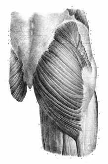
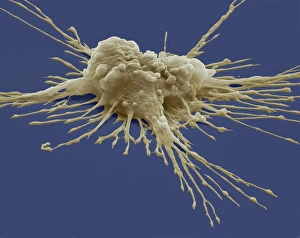
"Exploring the Intricacies of Medical Scan: Unveiling the Hidden Wonders Within" Delving into the depths of our brain, MRI scans reveal a mesmerizing world where normalcy resides. Witnessing the microscopic realm, SEM captures Lactobacillus casei bacteria in all its intricate glory. Peering into the mysteries of cognition, an MRI scan unveils the complexity and beauty of our brain's inner workings. Journey back in time with an 1866 arm bones anatomy engraving, showcasing the timeless elegance of human skeletal structure. Embark on a visual voyage through a normal abdomen with a stunning 3D CT scan that brings internal organs to life. Embracing technology's embrace, doctors utilize augmented reality to enhance their expertise and provide accurate diagnoses effortlessly. A glimpse into history reveals an 1866 back torso anatomy engraving, reminding us of our body's remarkable resilience and interconnectedness. Unlocking secrets from centuries ago, explore an 1866 pelvic femoral region anatomy engraving that showcases both strength and vulnerability within us all. Discovering hidden wonders beneath our skin, an 1866 frontal trunk anatomy engraving unravels layers upon layers of anatomical marvels waiting to be explored. The anterior knee region comes alive through an enchanting 1866 anatomy engraving—unveiling both grace and stability within this crucial joint complex.